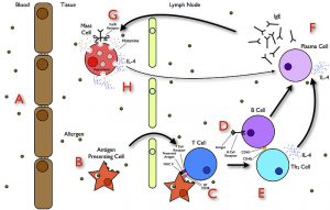
How does an allergy occur? The pathobiological mechanism involves several white blood cells that play a role in mounting an allergic reaction.
Table of Contents
When allergy season looms, some people with serious hypersensitivity to allergens tend to be apprehensive of what may come. Some would rather stay indoors than risking the odds of sucking up triggers that could instigate severe allergic reactions. Apart from triggers from the environment, other common factors for allergy include food, medication, certain toxins, venom from insect stings or bites, stress, and heredity. How does an allergy manifest? Which cells are involved in forming an allergic reaction?
The immune system
The immune system protects the body from foreign substances (generally referred to as antigens) that could pose a threat to our well-being. It prevents harmful bacteria, viruses, parasites, etc. from invading and causing harm. The white blood cells (also called leukocytes) constantly scout for antigens in order to destroy or disable them. The white blood cells include lymphocytes, neutrophils, basophils, eosinophils, monocytes, macrophages, mast cells, and dendritic cells.
Allergy – overview

An allergy is a state of hypersensitivity of the immune system in response to an allergen (i.e. a substance capable of inciting an allergic reaction). In this regard, several white blood cells play a role in mounting an allergic reaction.
In summary, the entry of an allergen into the body triggers an antigen-presenting cell, such as a dendritic cell. The dendritic cell takes up the allergen, process it, and then present its epitopes through its MHC II receptor on its cell surface. It, then, migrates to a nearby lymph node, waiting for a T lymphocyte to recognize it.
Upon recognition, the T lymphocyte may differentiate into a Th2 cell (type 2 helper T cells), which is capable of activating B lymphocyte. B lymphocyte, when activated, matures into a plasma cell that could synthesize and release IgE antibody in the bloodstream. Some of the circulating IgE may bind to mast cell and basophil. Thus, re-entry of such allergen could incite the IgE on mast cells and basophils to recognize its epitope. In effect, this activates the mast cell or basophil to release inflammatory substances (e.g. histamine, cytokines, proteases, chemotactic factors) into the bloodstream.
Anaphylaxis – a dreadful allergic reaction
The allergic reaction mounted by the immune system is supposed to protect the body. However, the allergens perceived by the body as a threat are generally harmless. The body tends to overly react to the allergens, and so leads to symptoms. Histamine, for instance, brings about the common symptoms of allergy: pain, heat, swelling, erythema, and itchiness.
Anaphylaxis is the most severe form of allergic reaction. It can occur rapidly and it affects more than one body system, such as respiratory, cardiovascular, cutaneous, and gastrointestinal systems. It occurs as a result of the release of inflammatory substances from mast cells and basophils upon exposure to an allergen. Within minutes to an hour, symptoms could manifest as a red rash, swelling, wheezing, lowered blood pressure, and in severe cases, anaphylactic shock.
In the presence of breathing difficulties, racing heart, weak pulse, and/or a change in voice, the situation is precarious. It calls for an immediate medical attention.
Why does anaphylaxis occur? IgE-mediated anaphylaxis is the common form of anaphylaxis. Initial exposure to an allergen leads to the release of IgE so that re-exposure to the allergen leads to its identification and the eventual activation of mast cells and basophils. Apart from immunologic factors, though, other causes of anaphylaxis are non-immunologic. For example, temperature (hot or cold), exercise, and vibration may cause anaphylaxis. In this case, IgE is not involved. Rather, these agents directly cause the mast cells and the basophils to degranulate.
Novel mechanism identified
Recently, a team of researchers1,2 found a novel mechanism that could explicate the hasty allergic reaction during anaphylaxis. They were first to uncover a mechanism involving the dendritic cells. Accordingly, a set of dendritic cells seem to “fish” allergens from the blood vessel using their dendrites. The dendritic cell near the blood vessel takes up the blood-borne allergen. Rather than initially processing it, and then presenting the epitope on its surface, it hands over the allergen inside a micro-vesicle to the adjacent mast cells.
Mast cells, unlike basophils that are in the bloodstream, are located in tissues, such as connective tissue. Thus, the question as to how the mast cells detect blood-borne allergen could be answered by the recent findings.
Rather than being internalized by the dendritic cells for processing, the allergen was merely taken into a micro-vesicle that budded off from the surface of dendritic cells. This, thus, saves time. It cuts the process, leading to a much rapid allergic reaction.
However, these findings were observed in mouse models. Therefore, the researchers have yet to observe if this novel mechanism also holds true on humans. If so, this could lead to possible therapeutic regulation of allergies, especially the most dreadful form, anaphylaxis.
— written by Maria Victoria Gonzaga
References
1 Choi, H.W., Suwanpradid, J. Il, Kim, H., Staats, H. F., Haniffa, M., MacLeod, A.S., & Abraham, S. N.. (2018). Perivascular dendritic cells elicit anaphylaxis by relaying allergens to mast cells via microvesicles. Science 362 (6415): eaao0666 DOI: 1126/science.aao0666
2 Duke University Medical Center. (2018, November 8). Using mice, researchers identify how allergic shock occurs so quickly: A newly identified immune cell mines the blood for allergens to directly trigger inflammation. ScienceDaily. Retrieved November 22, 2018 from www.sciencedaily.com/releases/2018/11/181108142440.htm

