Embryology
n., [ˌɛmbrɪˈɒlədʒɪ)]
Definition: The branch of biology that studies the formation and early development of living organisms.
Table of Contents
Embryology Definition
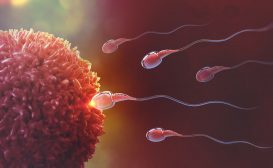
Embryology is a branch of biology that deals with the topics concerning gamete formation (gametogenesis), a fusion of gametes (fertilization), and embryo formation (embryogenesis). By definition, embryology encompasses the growth of both plant and animal embryos. But in a more general sense, we study animal or human embryology when referring to a medical sense.
Comparative Embryology
Comparative embryology is a sub-branch of embryology that deals with comparisons and differences between embryo developments of different species. This helps in developing clarity about how different animals are related to each other from evidence at the embryological developmental level.
Preformationism and epigenesis
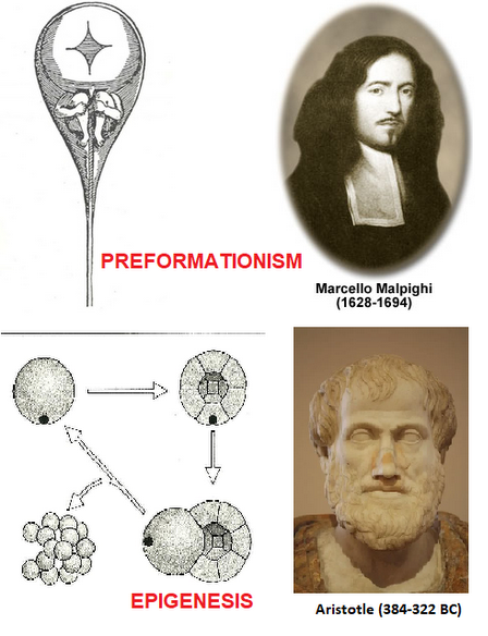
Two different theories are proposed for the early embryogenesis process. They are preformationism and epigenesis. Look at the differences below to understand the contrast.
Table 1: Preformationism and Epigenesis | ||
|---|---|---|
| Feature | Preformationism | Epigenesis |
| Proposed by | Marcello Malpighi | Aristotle |
| Basic idea | The development of organisms begins with pre-existing mini-versions or replicas. | The development of organisms is a sequential process that begins with gametes leading to an amorphous zygote. (Ref-1) |
| Embryonic structures are performed within sex cells/gametes | Yes | No |
| Embryonic structures arise newly after gamete fusion | No | Yes |
| Nature | Pre-assembled | Has to assemble (after fusion) |
| Idea of homunculus | Yes (homunculus unfolds into an adult) | No (sequential development from gametes to zygote to embryo to adult) |
| The current acceptance of the theory | Null; this theory has been disproven. | Accepted by the scientific community as the favored explanation for embryological development. |
Data source: Dr. Harpreet Narang of Biology Online
Embryology is the study of the embryo and its development from a single-celled zygote (fertilized ovum) to the establishment of form and shape (at which point, if it is an animal, it becomes a fetus). It is a subfield of developmental biology.
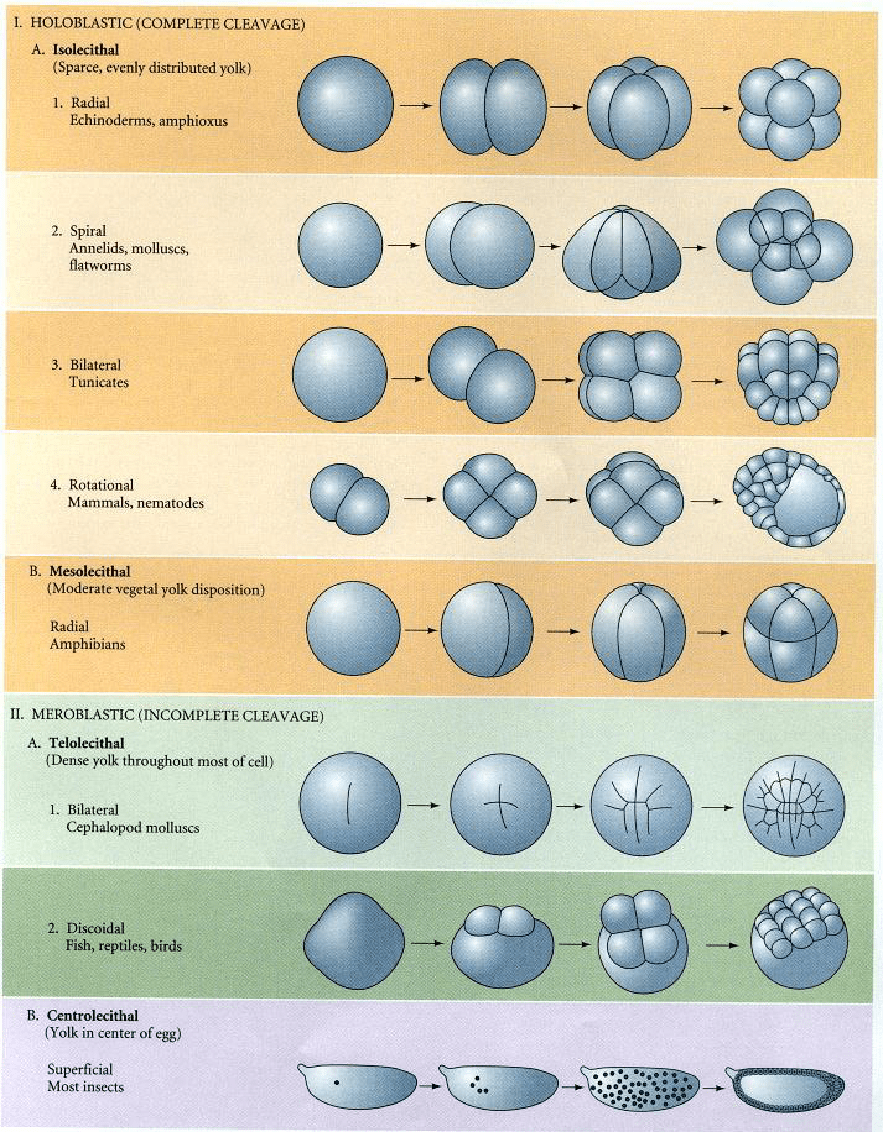
Holoblastic
Holoblastic cleavage is defined as the complete division of cells of the zygote and blastomeres.
Holoblastic cleavage leads to “doubling of the blastomeres” after each cleavage.
Four significant types of holoblastic cleavage in the isolecithal, mesolecithal cells, or microlecithal cells are:
- Bilateral (example: Tunicates)
- Radial (example: Echinoderms, Amphioxus, Amphibians)
- Rotational (example: Mammals, Nematodes)
- Spiral (example: Annelids, Molluscs, Flatworms)
The first stage/cleavage pattern in the holoblastic cleavage is always along the “vegetal-animal axis of the egg”.
The second cleavage pattern in the holoblastic cleavage is always “perpendicular” to the first cleavage pattern.
In holoblastic cleavage, the entire egg is involved in embryo formation, unlike meroblastic cleavage where some egg cells lead to yolk cell formation.
Meroblastic
Meroblastic cleavage is defined as the incomplete division of cells of the zygote and blastomeres.
Some cells of the blastomeres contribute to the yolk cell formation and thus don’t permit the protrusion of division furrow.
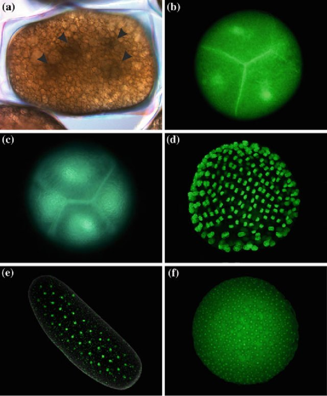
Three major types of meroblastic cleavage in the telolecithal and centrolecithal cells are:
- Bilateral (example: Cephalopod mollusks)
- Discoidal (example: Reptile, Fish, Birds)
- Superficial (example: Most insects)
Basal phyla
In basal phyla, animals have radial symmetry in their body plans. In the evolutionary process, these basal animals are believed to have segregated quite early on.
The four main basal phyla are:
Cleavage in basal phyla
- The cleavage pattern in the basal animals is “holoblastic radial cleavage”.
- This type of cleavage pattern in basal phyla results in radial symmetry.
- All cleavage divisions in the basal animal bodies rotate around a central axis during cleavage.
- These animals are also referred to as diploblastic as they have only one to two embryonic cell layers.

Bilaterians
Bilaterians are organisms that possess bilateral symmetry in embryonic stages.
Cleavage pattern: Can be either of the two — holoblastic or meroblastic; depending on the species
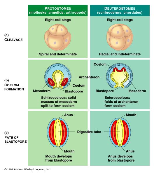
The animal kingdom is divided into two types based on the distinct character (embryological origins of the mouth and anus) during the gastrulation process.
- Blastopore becomes the mouth: Organisms are called protostomes. Examples: invertebrate organisms like insects, worms, and mollusks.
- Blastopore becomes the anus: Organisms are called deuterostomes. Example: vertebrate organisms like echinoderms, and chordates.
Germ layers
As embryonic development progresses, the blastula changes its forms into a more differentiated structure (gastrula). This process is called gastrulation and is accompanied by the formation of three distinct layers of cells called “germ layers”.
These germ layers are the precursors for all the bodily organs and tissues that develop later in the process from specialized cells.
- Endoderm: It is the innermost layer of cells.
Organs formed by endoderm: Digestive organs, nephrites, gills (or lungs or respiratory system), swim bladder, kidneys. - Mesoderm: It is the middle layer of cells.
Organs formed by intermediate mesoderm: Muscles, skeleton, blood organ system, endothelial cells, mesenchymal cells, cardiovascular system, reproductive system, gonadal ridge - Ectoderm: It is the outermost layer of cells.
Organs formed by ectoderm: Nervous system, brain, skin, hair, scales.
Drosophila melanogaster (fruit fly)
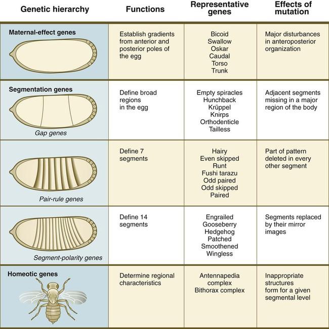
Drosophila melanogaster has been used as a model organism for different embryological studies. A number of genes have been described in fruit flies that play an integral role in embryo development. Some of them are:
- Maternal-effect genes
- Gap genes
- Pair-rule genes
- Segment-polarity genes
- Homeotic (Hox) genes
Humans
- Human beings are classified as bilateral animals.
- They display holoblastic rotational cleavage.
- Also classified as deuterostomes.
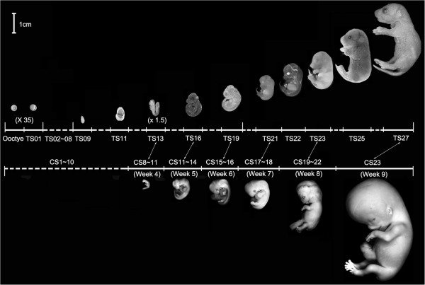
- The human embryo is defined as a dividing ball of cells starting from the zygotic stage, through the implantation stages (fourth week), and up till the end of the 8th week after conception.
- A human fetus (developing fetus) is defined as the developing stage beyond the 8th week after conception.
Evolutionary Embryology
Evolutionary embryology is studied as an ‘expanded field of comparative embryology’.
The theory of evolution proposed by Charles Darwin brought about a reorganization of comparative embryology and redirected its attention toward evolutionary perspectives.
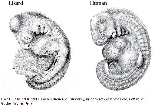
In 1842, Charles Darwin came across Johannes Müller’s summary of von Baer’s laws. At this point, he realized that “similarities observed during embryonic development” could serve as “compelling evidence in support of the genetic relationships between various animal groups”.
Darwin famously stated in his 1859 work, On the Origin of Species, that “a community of embryonic structure reveals community of descent”.
Even Karl Ernst von Baer’s principles explained the same idea. It emphasized why the diversity of species often has similarities in the appearance of early developmental stages.
Importance of evolutionary embryology: Understanding a species’ evolution can be enhanced by studying embryology because certain homologous structures are exclusively observable during the embryonic phase.
This is illustrated by the presence of a tail in the early development of all vertebrate embryos, including those of humans, chickens, and fish, even if the fully developed organism lacks a tail.
Von Baer’s principles
The four most important Von Baer’s principles are:
- The basic characteristics tend to emerge earlier during the developmental process compared to the specific features.
- The more specific characteristics typically arise from the more general ones during development.
- The embryo of a particular species never bears any resemblance to the fully-grown form of a different, lower species.
- The embryo of a particular species may share some resemblance to the embryonic form of a lower species.

Origins of Modern Embryology
Modern embryology began after the mammalian ovum studies undertaken by Karl Ernst von Baer in 1827.
It is believed that the development of ultrasound technology in the late 1950s led to a deeper embryology understanding.
Germ layer theory: Proposed by Karl Ernst von Baer and Heinz Christian Pander. This theory helped in understanding the progressive steps of embryo development. This theory was also important because it helped in understanding why embryos of different organisms often share similarities during the early developmental stages.

Modern embryology research
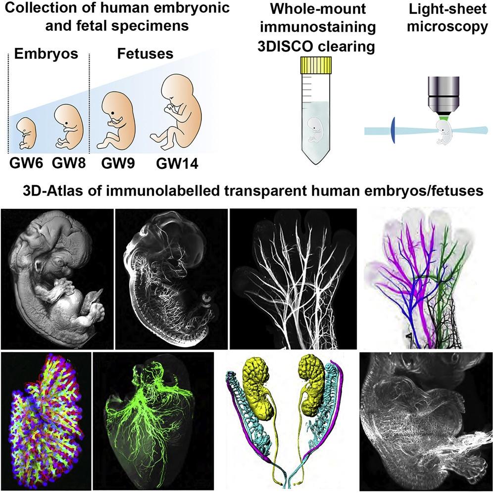
Modern embryology research revolves around visionary goals ranging from understanding the genetic control of the embryo development processes, embryonic stem cell research, gestational surrogacy, and studies of donor cells in IVFs to nuclear transfer studies and their applications in human therapeutic medicine for affected individuals. For instance, the activation of transcription factors and many other orchestrated biological events were found to be crucial during embryonic development. In particular, they drive the stem cells to form mature cell types that ultimately build an entire organism. (Ref. 8)
Medical Embryology
- Widely used for the detection of fetal development abnormalities and birth defects.
- Widely used for the exploration of modern tools and strategies to deal with these abnormalities.
- Widely used for dealing with genetically derived malformations.
- Widely used for effectively dealing with birth syndromes.
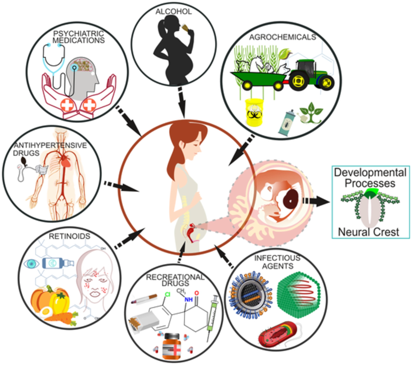
- Widely used for dealing with disruptions in embryos caused by external factors called “teratogens”. Some examples of teratogens are:
- Alcohol
- Retinoic acid
- Ionizing radiation
- Hyperthermic stress
Vertebrate and Invertebrate Embryology
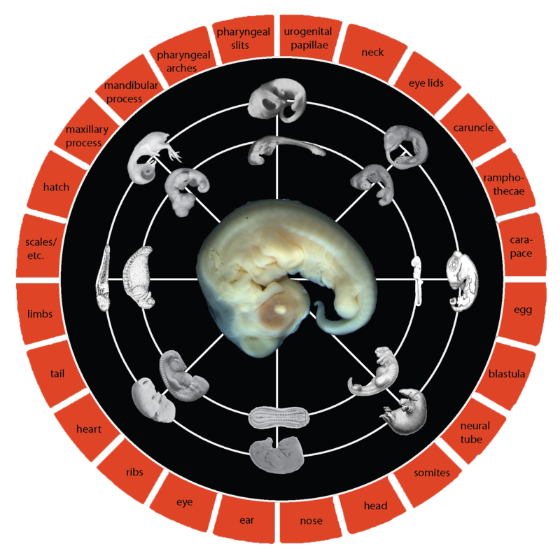
There are many common theories that apply to both vertebrate and invertebrate embryology. Within invertebrates, there are some variations specific to a species’ embryology. For this reason and to make comparisons easier among different species, a character-based system has been developed. It is called the “Standard Event System”. This system documents all the variations and permits phylogenetic comparisons among species.
Birth of Developmental Biology
Developmental biology works in conjunction with molecular biology, genetics, and morphology. Using molecular tools, developmental biologists study the correlation of developmental genes with morphological change. This helps them to understand which and how genes regulate the morphological development in an embryo.
History
Let’s look at the understanding of embryology from a historical perspective.
Ancient Egypt
Ancient Egyptians are known to view the placenta as the seat of the soul. The knowledge about embryo development from an egg (female gamete) was stated by one of the old Egyptian literature from Akhenaten times.
Ancient India
Ancient Indians are also known to possess vast knowledge of embryology. While the Asian population had many prevailing notions, the ones from India seem more advanced and better elucidated. Several ancient Hindu literatures like Bhagavad Gita, Sushruta Samhita, and Bhagavata Purana tell about a deeper understanding of the embryology subject from ancient India. The age-old medical tradition of India popularly known as Ayurveda too had some understanding of human embryology.
Ancient Greece
Greek embryology dates back to pre-Socratic philosophers, who had opinions on various aspects of embryology. Some of the most well-known early ideas on embryology come from Hippocrates, who claimed that the development of the embryo is put into motion by fire and that nourishment comes from food and breath introduced into the mother.
Aristotle‘s views were similar to Hippocrates, and he believed that the male had a seed that developed into the embryo within the female womb. Epicureans also claimed amniotic fluid nourishes the fetus. Herophilus talked about the ovaries and fallopian tubes in his studies. Greek discussions on embryology often tried to answer several questions, such as the origin of the seed and whether both the male and the female had a seed that made a contribution to the developing embryo.
Patristics
Patristic authors discussed embryology, including the value of the fetus and when it acquires value. Some authors believed the soul was present at conception, while others thought it entered at a later stage. They also discussed the relationship between the soul and the embryo, with three possible conceptions: the soul pre-exists and enters at conception, the soul enters existence at conception, or the soul enters the human body after it forms.
These ideas were debated among different Christian traditions, including Nestorian, Monophysite, and Chalcedonian. Later, discussions focused on synhyparxis, the idea that the soul is present at conception.
Embryology in Jewish tradition
Many Jewish authors discussed embryology, particularly in relation to the impurity of the mother after childbirth, in the Talmud.
The embryo is described as developing through various stages, including golem, shefir meruqqam, ubbar, walad, walad shel qayama, and ben she-kallu khadashaw.
The Talmud sages believed that both the male and female contribute to the formation of the fetus, with the relative proportions of their seeds determining the sex of the child.
Some Talmudic texts discuss magical influences on the development of the embryo, such as sleeping on a bed pointed to the north–south to have a male child.
Talmudic embryology draws on Greek discourses, particularly from Hippocrates and Aristotle, but also offers some novel statements on the subject.
There are notions of male embryos and female embryos.
You might like to read: Sex Determination in Humans and the sex-determining region Y gene.
Embryology in the Islamic tradition
The Qur’an describes the development of an embryo in four stages, similar to Galen’s description. This knowledge was transmitted through translations of Greek medical texts by figures such as Sergius of Reshaina and the Academy of Gondishapur. Islamic legal tradition also includes discussions on embryology. Similar descriptions are also found in the Syriac Jacob of Serugh’s letter to Archdeacon Mar Julian.
“The Science behind Embryonic Cleavage”
The idea of cleavage developed during the 19th century when biologists and physicians started using the newly developed instrument called the microscope. This helped in elucidating the sequential formation of an embryo from a zygote. This shed light on the epigenesis theory and disproved preformationism.
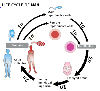
Cleavage encompasses all the initial steps of embryo development. It is accompanied by sequential mitotic divisions that follow fertilization (fusion of male [sperm] and female [egg] gametes). Cleavage is specific for each organism (species).
Based on the extent of the division of cells, there are two types; complete and incomplete. Complete division of cells is called “holoblastic cleavage” whereas an incomplete division of cells is called “meroblastic cleavage”.
Watch this vid to learn more about embryology:
Take the Embryology Quiz!
Further Reading
- Biology Tutorial – Developmental Biology
References
- Van Speybroeck, L., De Waele, D., & Van de Vijver, G. (2002). Theories in early embryology: close connections between epigenesis, preformationism, and self-organization. Annals of the New York Academy of Sciences, 981, 7–49.
- Maienschein, Jane, “Epigenesis and Preformationism”, The Stanford Encyclopedia of Philosophy (Spring 2017 Edition), Edward N. Zalta (ed.)
- Iyer, S., Mukherjee, S., & Kumar, M. (2021). Watching the embryo: Evolution of the microscope for the study of embryogenesis. BioEssays : news and reviews in molecular, cellular and developmental biology, 43(6), e2000238. https://doi.org/10.1002/bies.202000238
- Belle, M., Godefroy, D., Couly, G., Malone, S. A., Collier, F., Giacobini, P., & Chédotal, A. (2017). Tridimensional Visualization and Analysis of Early Human Development. Cell, 169(1), 161–173.e12. https://doi.org/10.1016/j.cell.2017.03.008
- Molecular Basis for Embryonic Development https://clinicalgate.com/molecular-basis-for-embryonic-development/
- Xue, L., Cai, J. Y., Ma, J., Huang, Z., Guo, M. X., Fu, L. Z., Shi, Y. B., & Li, W. X. (2013). Global expression profiling reveals genetic programs underlying the developmental divergence between mouse and human embryogenesis. BMC genomics, 14, 568. https://doi.org/10.1186/1471-2164-14-568
- Cerrizuela, S., Vega-Lopez, G. A., & Aybar, M. J. (2020). The role of teratogens in neural crest development. Birth defects research, 112(8), 584–632. https://doi.org/10.1002/bdr2.1644
- Huilgol, D., Venkataramani, P., Nandi, S., & Bhattacharjee, S. (2019). Transcription Factors That Govern Development and Disease: An Achilles Heel in Cancer. Genes, 10(10), 794. https://doi.org/10.3390/genes10100794
©BiologyOnline.com. Content provided and moderated by Biology Online Editors.

