Antecubital fossa
n., plural: antecubital fossae
[an″tĭ-ku´bĭ-t’l ˈfɒsə]
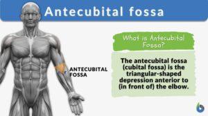
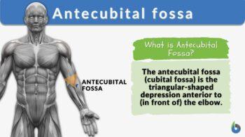
Definition: the depression anterior to (in front of) the elbow
Table of Contents
Antecubital Fossa Definition
The antecubital fossa or the cubital fossa is the triangular-shaped hollow depression between the forearm and the anatomical arm. The antecubital fossa is on the anterior side of the elbow that has a distal apex. This site is commonly used for venous and arterial cannulation for drawing a blood sample.
What is the antecubital?
- Meaning of antecubital: of or relating to the crook of the elbow
- Define fossa: Fossa is a shallow depression or hollow space or groove
Watch this vid about antecubital fossa:
Biology definition:
The antecubital fossa is the fossa in the anterior of the cubitus or simply the depression in front of the elbow. It contains the tendon of the biceps, the median nerve, and the brachial artery.
It is the region where blood is commonly drawn from since superficial veins cross through it. It is the site where blood pressure is measured. The stethoscope is placed over the brachial artery in the fossa. It is also an area used to palpate the brachial pulse.
Etymology: antecubital, from Latin ante, meaning “before”, “in front of”, “opposite”, Latin “cubitum”, meaning “elbow”; fossa, from Latin “fossus”, meaning “dug up”
Variant: anticubital fossa
Synonyms: cubital fossa; fossa cubitalis; chelidon; triangle of elbow
Where Is the Antecubital Fossa?
Where do we find the antecubital fossa? Why is it called the antecubital? Anatomically, the antecubital fossa is the transitional triangular depression or hollow area located on the anterior side of the elbow (or Latin cubitus, the medical term for elbow), between the forearm and the anatomical arm. This is why it is called the antecubital.
The antecubital fossa, also referred to simply as the cubital fossa, is the connecting portion of the forearm’s ulna with the humerus and the radius. (Figure 1)
The synovial joint between the arm bones along with several muscles and tendons creates the antecubital fossa. On the top side, skin is present along with nerves and muscles while at the bottom are the muscles.
This site is widely used for venous and arterial cannulation for drawing blood samples.

Note it!
Fossa Definition in Anatomy: Fossa is the groove or depression area in the body.
Examples:
- The middle fossa is the depression in the skull base which seems to have a butterfly shape.
- The triangular fossa is the groove on the top part of the ear auricle.
Antecubital Region
The antecubital region of the body is a triangular region on the anterior side of the elbow between the forearm and the anatomical arm. The antecubital fossa on the right arm is known as the right antecubital and the antecubital fossa on the left arm is known as the left antecubital.
Antecubital Area
The antecubital area of the arm or the triangular region of the antecubital fossa has three borders namely, superior, medial, and lateral borders.
- Superior border – Imaginary horizontal line in brachioradialis muscle
- Medial border – lateral border of the muscle pronator teres
- Lateral border – medial border of the muscle brachioradialis
Apart from the three borders, the antecubital fossa has a roof and a floor which are formed by:
- Roof: Subcutaneous fat and skin along with fascia and bicipital aponeurosis (a tendon-like sheet that arises from the biceps brachii tendons) form the roof. The bicipital aponeurosis is the protective covering over the ulnar artery, brachial artery, and medial nerve. The median cubital vein, which is punctured for blood sample collection, runs through the roof of the antecubital fossa.
- Floor: Supinator from distal side and brachialis from proximal side forms the floor.
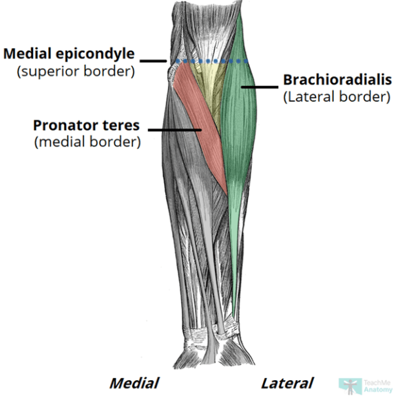
Antecubital Space
- The narrow triangular space formed by three muscles along with a roof and floor on the anterior side of the elbow joint is the antecubital fossa space.
- This antecubital fossa space provides the home to a number of critically important nerves and blood vessels.
- Also, several muscles and tendons cross through the antecubital fossa space, which is essential for the normal functioning of the arm or the upper limb.
Note it!
Is antecubital fossa a vein? No, antecubital fossa is not a vein. However, it is the cavity in the anterior surface /region of the elbow from where blood samples are withdrawn as it houses three antecubital fossa veins or blood vessels used for drawing blood for diagnostic purposes.
Antecubital Fossa Anatomy
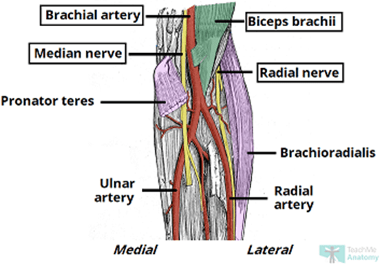
A figurative representation of the antecubital fossa is shown in Figure 3.

Radial Nerve
The radial nerve passes through the vicinity of the antecubital fossa below the brachioradialis muscle; it is essentially not an integral part of the antecubital fossa. The radial nerve divides here into two, i.e., superficial and deep branches.
The deep branch is involved in the motor function and is connected to the posterior part of the forearm whereas the superficial branch is involved in the sensory functions and runs deep into the brachioradialis muscle. Thus, both deep and superficial branches are critical for the normal functioning of the forearm.
Biceps Tendon
Bicep tendons are connected to the radius bone resulting in the formation of a prominent structure of antecubital fossa, referred to as radius tuberosity.
The bicep tendon passes through the center, distally to the neck of the radius in the antecubital fossa. It also forms the bicipital aponeurosis which is part of the roof of the antecubital fossa.
Brachial Artery
The brachial artery is the oxygen-supplying artery of the forearm that divides into radial and ulnar arteries at the antecubital fossa apex. The brachial artery is located between the median nerve and the biceps tendon.
The pulse of the Brachial artery can be felt by palpating the medial to the biceps tendon in the antecubital fossa. The brachial artery can also be used in coronary or peripheral angiography, especially in cases wherein the patient can not lie flat.
Median Nerve
The median nerve innervates the antecubital fossa medially and exits the antecubital fossa via two pronator teres heads, i.e., ulnar and humeral heads. This nerve has no branches in the arm or axillae however its articular branch supply to the elbow joint.
Blood Supply in the Antecubital Fossa
The major blood supply of the antecubital fossa includes the brachial artery and two superficial antecubital veins. The brachial artery divides into two at the distal apex of the antecubital fossa i.e., ulnar and radial arteries. These arteries run through the forearm and supply to the anterior as well as the posterior part of the arm. These arteries run through deep as well as superficial areas of the hand.
Under the roof of the cubital fossa, two superficial veins (cephalic veins and basilic veins) connect with the median cubital vein. The right or left cephalic vein starts in the wrist from the anteromedial region while the basilic vein begins in the wrist at the anterolateral surface.
The median cubital vein connects with two veins i.e., cephalic veins and basilic veins on its way to the forearm. The basilic vein further connects with the brachial vein to form an axillary vein at the lower border of the teres major. While the cephalic vein and the basilic veins combine to drain out the contents into the axillary vein in the axilla region.
Antecubital Fossa Nerves
The antecubital fossa has two primary nerve supplies, namely, median and radial nerves.
- Median nerves – These nerves have roots in C5-T1 and supply to the posterior part of the forearm. These nerves have a role in the movement of fingers and extension of the wrist.
Also, these nerves provide sensory nerve supply to the dorsal side of the lateral 3 & 1/2 digits and dorsum of the hand. - Radial nerves – These nerves have roots in C6-T1 and supply primarily to the anterior forearm muscles, hand muscle of thenar eminence, and the two medial lumbricals. These nerves have a role in forearm pronation, flexing of the wrist, and finger movement. These nerves also provide the sensory nerve supply to the volar surface of the lateral 3 & 1/2 digits and lateral palm.
- Median nerves – These nerves have roots in C5-T1 and supply to the posterior part of the forearm. These nerves have a role in the movement of fingers and extension of the wrist.
Antecubital Fossa Muscles
Four muscles form the essential part of the antecubital fossa, namely:
- Pronator teres
- Median border of the fossa is the Probator teres. Pronator teres muscle has two heads namely,
- Ulnar head, originates from the coronoid process of the ulna
- Humeral head that originates from the medial epicondyle
- Median nerves innervate the pronator teres and pass through the two heads of the pronator teres.
- Median border of the fossa is the Probator teres. Pronator teres muscle has two heads namely,
- Brachioradialis
- It is the lateral border of the antecubital fossa. Radial nerve supplies to the brachioradialis.
- Brachioradialis helps to flex the forearm.
- The brachioradialis starts from the lateral supracondylar ridge of the humerus and connects to the distal radius.
- Brachialis
- The brachialis is the proximal part of the floor and the musculocutaneous nerve supply to it.
- It starts from the shaft of the humerus and goes inside the radial tuberosity of the ulna.
- Supinator
- Supinator forms the distal part of the floor and posterior interosseus branch of the radial nerve.
- Supinator has two heads — one that originates from the lateral humeral epicondyle and the second head originates from the posterior ulna. These two heads combine and get inside the posterior radius.
- Supinator helps in supination of the forearm.
- Pronator teres
Physiological Variations
According to the presence of the median cubital vein (MCV) or median antebrachial vein, the superficial veins of the Antecubital fossa can be classified into the following four types:
- Type I: Also, called N type. Herein, the dominant vein is median antebrachial, and in the cubital region, it combines with both basilic and cephalic veins.
- Type II: Also, called M type. Herein, in the cubital region, the median cubital vein joins with both the cephalic vein and basilic vein.
- Type III: Herein, the brachial cephalic vein is absent or faintly present in the cubital region.
- Type IV: Herein, any connection between the cephalic vein and basilic vein is missing.
Type II is most commonly found, followed by Type I.
Clinical Significance of the Antecubital Fossa
- The venipuncture for taking the blood sample is carried out in the median cubital vein which is present superficially on the roof of the cubital fossa.
- The stethoscope is placed over the cubital fossa to measure the blood pressure by hearing the Korotkoff sounds in the brachial artery.
- Cubital tunnel syndrome wherein, in the cubital fossa, an ulnar neuropathy occurs. In this syndrome, the ulnar nerve becomes compressed within the cubital tunnel. The patient experiences sensory paraesthesia in the ulnar distribution of the hand. The patient exhibits motor symptoms like clumsiness in the intrinsic hand movement.
- Supracondylar Fracture occurs due to a fall with a hyper-extended elbow which causes a transverse fracture. The resultant ischemia may further cause Volkmann’s ischaemic contracture.
Answer the quiz below to check what you have learned so far about antecubital fossa.
References
- Bains, K.N.S., Lappin, S.L.(2021). Anatomy, Shoulder and Upper Limb, Elbow Cubital Fossa. [Updated 2021 Jul 26]. In: StatPearls [Internet]. Treasure Island (FL): StatPearls Publishing; https://www.ncbi.nlm.nih.gov/books/NBK459250/
- Ellis, H., Feldman, S., & Harrup-Griffiths, W. (2010). The clinical anatomy of the antecubital fossa. British journal of hospital medicine (London, England : 2005), 71(1), M4–M5. https://doi.org/10.12968/hmed.2010.71.Sup1.45982
- Lee, H., Lee, S. H., Kim, S. J., Choi, W. I., Lee, J. H., & Choi, I. J. (2015). Variations of the cubital superficial vein investigated by using the intravenous illuminator. Anatomy & cell biology, 48(1), 62–65. https://doi.org/10.5115/acb.2015.48.1.62
- Sheen, J.R., Khan, Y.S. (2021). Anatomy, Shoulder and Upper Limb, Cubital Fossa. [Updated 2021 Jul 27]. In: StatPearls [Internet]. Treasure Island (FL): StatPearls https://www.ncbi.nlm.nih.gov/books/NBK551674/
©BiologyOnline.com. Content provided and moderated by Biology Online Editors.

