ˈak.son ˈtɚmɪnəl
The axon terminal is important in cell to cell communication through the neurotransmitters it releases into the synaptic cleft. The neurotransmitters that exit the neuron relay signals to the next target cell.
Table of Contents
An axon terminal is any of the button-like endings of axons through which axons make synaptic contacts with other nerve cells or with effector cells. At the axon terminal, synaptic vesicles containing neurotransmitters are docked. Upon activation by a graded potential or by an action potential of the presynaptic neuron, the cell allows the entry of calcium ions. This triggers a cascade reaction resulting in the synaptic vesicles fusing to the membrane of the axon terminal. The synaptic vesicle proteins that are activated by the calcium ions form fusion pores through which the neurotransmitters can exit. The neurotransmitters exert their effects on the target cell within a limited span of time. Soon after, the neurotransmitters are absorbed back to the presynaptic neuron or degraded metabolically by enzymes. The axon terminal is therefore essential in cell to cell communication. It is crucial in providing a means for neurotransmitters to exit the neuron and relay signals to the target cell.
Axon Terminal Definition
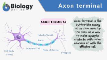
An axon terminal refers to the axon endings that are somewhat enlarged and often club- or button-shaped. Axon terminals are that part of a nerve cell that make synaptic connections with another nerve cell or with an effector cell (e.g. muscle cell or gland cell).
Etymology
The term axon came from the Ancient Greek ἄξων, meaning “áxōn” or “axis”. The term terminal is from Latin terminalis, which pertain to a boundary or to an end. It came from Latin terminus, meaning “a limit” or “an end”. Synonyms: axon terminal bouton; bouton terminaux; pieds terminaux; synaptic bouton; synaptic knob; synaptic ending; synaptic terminal; terminal bouton.
Neuronal structure
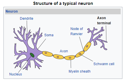
A neuron (also called a nerve cell) is an excitable cell in the body of higher animals, including humans. The cell can be distinguished from other cell types in having distinctive parts, such as soma, dendrites and axons.
- The soma is the cell body that contains the organelles, such as the nucleus.
- Both dendrites and axons are cytoplasmic processes. While dendrites are the branched threadlike projections, the axon is a single, slender, longer fiber of a neuron. It is also called a nerve fiber. The function of the axon is to carry efferent (outgoing) action potentials and conduct nerve impulse away from the cell body to a synapse. Conversely, the dendrites receive the nerve impulse from another neuron (via a synapse), and then propagate the electrochemical stimulation to the cell body. At the end of an axon, there is a so-called axon terminal that is button-like and is responsible for providing synapses between neurons. The axon terminal contains specialized chemicals called neurotransmitters that are initially contained inside the synaptic vesicles. In humans, the axon can be over a foot long. In the peripheral nervous system, the larger (myelinated) axons are surrounded by a myelin sheath formed by concentric layers of the plasma membrane of the Schwann cell. An axon hillock is a tapering region between the cell body and its axon. This region is responsible for summating the graded inputs from the dendrites and producing action potentials if the threshold is exceeded.
Axon terminal and synapse
An axon terminal contains various neurotransmitters that are released at the small gap between two communicating neurons. This gap is called a synapse. The neuron that sends nerve impulses by releasing neurotransmitters via the axon terminal at the synapse is called a presynaptic neuron. In contrast, the neuron that receives the impulse is called a postsynaptic neuron.
The synapse that serves as the junction between two neurons may be of two types: a chemical synapse or an electrical synapse.
- A chemical synapse is one that is involved in the transmission of nerve impulses from a neuron to another neuron or from a neuron to an effector cell (e.g. a muscle cell or a gland cell).
- An electrical synapse is a junction between two neurons that are in juxtaposition. This type of synapse provides faster nerve impulse transmission.
Synaptic activity
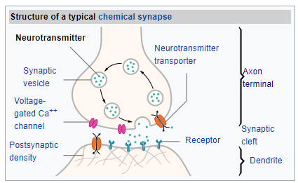
Neurons transmit nerve impulses by means of electrochemical signals and neurotransmitters. Neurotransmitters belong to a group of chemicals that are released on cue. Neurotransmitters are stored in synaptic vesicles. These vesicles are located in the axon terminal of a presynaptic neuron of the central or peripheral nervous system. There are many types of neurotransmitters and they may be either excitatory or inhibitory.
Examples of neurotransmitters are acetylcholine, noradrenaline, adrenaline, dopamine, glycine, y aminobutyrate, glutamic acid, substance P, encephalins, endorphins, and serotonin. These chemicals are responsible for relaying signals from a neuron to the target cell across a synapse. They are kept inside the synaptic vesicles. The vesicles then move down the axon and finally to the axon terminal where they will be clustered near the plasma membrane.
The transmission of nerve impulses begins with a graded electrical potential or with an action potential traveling along the membrane of the presynaptic neuron until the synapse. Electrical depolarization of the membrane at the synapse will lead to increased permeability to calcium ions. An influx of calcium ions activates calcium-sensitive proteins into releasing the neurotransmitters into the synaptic cleft.
The release of neurotransmitters is by exocytosis. The calcium ions binding to the synaptic vesicle proteins results eventually in the fusing of secretory vesicles into the presynaptic membrane and a formation of a fusion pore as the vesicle proteins move apart. The transient pore provides a means for the release of intravesicular contents from the presynaptic neuron into the synaptic cleft. After the secretion, the pore is eventually sealed.
These neurotransmitters bind to the receptors of the target cell. If the target cell is another neuron, the neurotransmitters bind to the postsynaptic receptors on the dendritic membrane of the postsynaptic neuron. The neurotransmitters may act as either excitatory or inhibitory. An example is when a neuron receives more excitation than inhibition from the neurons connected to it, the neuron will be activated to generate a new action potential at its axon hillock to signal the release of neurotransmitters that will relay the impulse to the new target cell (e.g. another neuron).
The neurotransmitters in the synaptic cleft are often available for only a short period of time. Those that did not bind to the post-synaptic receptors and therefore have not been used for synaptic transmission, their possible fates are as follows: (1) reuptake by the presynaptic neuron, (2) metabolic degradation by enzymes.
Importance
The axon terminal is the part of the axon that releases the neurotransmitters that relay signals across a synapse. For example, in a neuromuscular junction, the axon terminal releases neurotransmitters to relay nerve impulses from the neuron to the target cell, which may be another neuron, a muscle cell, or a gland cell.
Acetylcholine, for example, is the neurotransmitter that is released by a neuron to stimulate a muscle cell. If the axon terminal fails to release acetylcholine, there will be no chemical to activate the target muscle. As such, this could lead to paralysis.
Apart from acetylcholine, other major neurotransmitters that the axon terminal provides a way across the synapse are dopamine, norepinephrine, epinephrine, serotonin, oxytocin, somatostatin, etc. These chemicals are important chemical regulators of many biological activities. Without an axon terminal to mediate their release from the neuron, these chemicals, similar to acetylcholine, will not be able to exert their crucial role. Thus, the communication between cells will be obstructed by a dysfunctional axon terminal. Apart from that, the reuptake of unused neurotransmitters from the synaptic cleft back to the presynaptic neuron would be affected if there is no functional axon terminal.
Try to answer the quiz below to check what you have learned so far about axon terminals.
References
- The Neuron. (2009). Retrieved from Ship.edu website: http://webspace.ship.edu/cgboer/theneuron.html
- 16.2 How Neurons Communicate – Concepts of Biology – 1st Canadian Edition. (2019, May). Retrieved from Opentextbc.ca website: https://opentextbc.ca/biology/chapter/16-2-how-neurons-communicate/
- Neurons, Synapses, Action Potentials, and Neurotransmission – The Mind Project. (2020). Retrieved February 24, 2020, from Ilstu.edu website: http://www.mind.ilstu.edu/curriculum/neurons_intro/neurons_intro.php
- Acetylcholine Neurotransmission (Section 1, Chapter 11) Neuroscience Online: An Electronic Textbook for the Neurosciences Department of Neurobiology and Anatomy – The University of Texas Medical School at Houston. (2019). Retrieved from Tmc.edu website: https://nba.uth.tmc.edu/neuroscience/m/s1/chapter11.html
- Calcium Influx: Initiation of Neurotransmitter Release. (2019). Retrieved from Williams.edu website: https://web.williams.edu/imput/synapse/pages/IIA1.htm
© Biology Online. Content provided and moderated by Biology Online Editors

