Cell morphology
n., plural: cell morphologies
[sɛl mɔɹˈfɑlədʒi]
Definition: the form, shape, size of a cell
Table of Contents
The basic essence for any living organism is its structural framework which includes appearance, form, and the visually-recognizable characters. This is what’s called the morphology of that organism. The morphological branch of biology deals with the very basic size, form, shape, structure, color, texture, and pattern that a biological being possesses. Morphology can be studied at various different levels like organismal, organ, tissue, or cellular morphology. So, the definition of cell morphology in biology is related to the morphological and phenotypic description at the cellular level. Cell morphology is the topic of concern here so let’s proceed with it and try to understand what it is, what role it serves, how many types of morphology of cells occur and its spectrum across various lineages of life…
Cell Morphology Definition
Cells are the most basic and vital unit of all living/biological organisms. Cell morphology entails the structural framework of this basic unit of life. It encompasses the various shapes and sizes in which cells make up a living being. So, if one is asked to define cell morphology, it’s the concept that deals with all the possible “structural manifestations of cells” whether it be in prokaryotes or eukaryotes. It, in totality, comprises all the possible cell shapes and cell sizes.
Biology definition:
Cell morphology pertains to the morphological features of the cell(s). It employs microscopy to identify the shape, structure, form, color, texture, pattern, and size of the cell(s). In bacteriology, for instance, cell morphology pertains to the shape of bacteria if cocci, bacilli, spiral, etc., and the size of bacteria. Thus, determining cell morphology is essential in bacterial taxonomy.
Synonyms: cellular morphology; cytomorphology.
Definition of Cell Morphology in Microbiology
What is cell morphology in microbiology? In microbiology, cell morphology is the branch of science dealing with the cell’s structure, shape, size, and arrangement of prokaryotes. Prokaryotes are the basic form of living organisms that are devoid of a well-defined nucleus, instead have a nucleoid, plus have no specialized organelles as eukaryotes. These organisms are typically unicellular and their morphology is studied under the domain of prokaryotic microbiology. Both eubacteria and archaea come under prokaryotes. Now since bacteria are unicellular, the cell shape defines the bacteria’s shape. Just like humans come in different shapes and sizes, so do bacteria. Let’s take a look at the different types of cell morphology of bacteria.
Types of Bacterial cell morphology
Below are the different bacterial cell morphologies:
- Coccus (plural cocci): Spherical shape, like tiny balls
- Bacillus (plural bacilli): Rod shape, like cylinders
- Spiral: twisted like a DNA helix
- Vibrio: comma-shaped
The bacterial taxonomy is representative of the bacterial cell morphology most of the time. The genus name usually carries an identifier to the bacterial shape like Streptococcus has “coccus” in it, meaning it’s spherical in shape. Helicobacter has “helico” in its name, meaning its spiral/helical shape. Based on the cell morphology and arrangement, there are further classifications within these 4 major types like for pairs (diplo- prefix used), chains (strepto- prefix used), etc.
Cell Morphology Examples in Prokaryotes
Look at the table and figures below depicting the different cell morphologies in prokaryotes.
Table 1: Different Cell Morphologies in Prokaryotes | |
|---|---|
| Cell Morphology | Examples of Prokaryotes |
| Coccus | Staphylococcus spp., Streptococcus spp., Micrococcus spp., Neisseria spp., Gonococcus, Sarcina spp., Moraxella spp., Enterococcus spp. |
| Bacillus | Bacillus spp., Clostridium spp., Escherichia spp., Klebsiella spp., Pseudomonas spp., Proteus spp., Salmonella spp., Actinomyces spp. |
| Spiral | Spirillum spp., Campylobacter spp., Helicobacter spp., Leptospira spp., Borrelia spp., Treponema spp. |
| Vibrio | Vibrio cholerae, Vibrio parahaemolyticus, Vibrio vulnificus, Vibrio gastroenteritis, Vibrio harveyi |
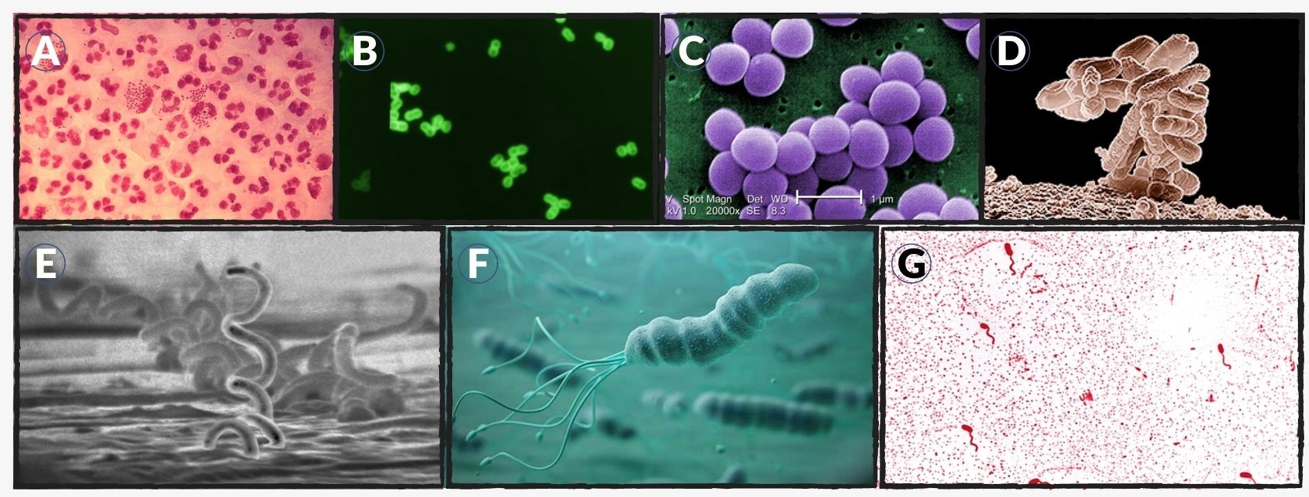
Figure 1: Cell morphology of different bacteria:
- (A) Neisseria gonorrhoeae or the Gonococcus is a gram-negative coccus-shaped bacterium. It causes gonorrhea, a sexually-transmitted disease. Credit: CDC/ Joe Millar.
- (B) Streptococcus pneumoniae is a gram-positive coccus-shaped bacterium. It is one of the main causal organisms of pneumonia. Credit: CDC/ Dr. M.S. Mitchell.
- (C) Staphylococcus aureus is a gram-positive coccus-shaped bacterium. It, along with Streptococcus pyogenes, causes the toxic shock syndrome. Credit: CDC/ Matthew J. Arduino, DRPH.
- (D) Escherichia coli, the model organism used for bacterial studies is a rod-shaped bacterium. Credit: Eric Erbe/USDA.
- (E) Treponema pallidum is a spiral bacterium that causes syphilis. Credit: CDC/ Dr. David Cox.
- (F) Helicobacter pylori is a spiral bacterium that causes peptic ulcers by damaging the duodenum in the digestive system. Credit: John Hopkins Medicine.
- (G) Vibrio cholerae causes cholera- an intestinal infection. Source: Denniss, public domain.
Cell Morphology Examples in Eukaryotes
Eukaryotes are organisms that have a well-defined nucleus and specialized organelles in their cells. They can be both unicellular or multicellular. Hence, it’s not necessary that the cell morphology of eukaryotic cells will define the eukaryotic organism’s morphology.
In the medical context, cell morphology is the phenotypic presentation of a cell tightly linked with its role in a eukaryote’s body. In eukaryotes, mostly all organisms are multicellular. The cells are in synchrony with each other to make the body functional. Hence, a hierarchical system is as follows: cells » tissues » organs. Different tissues of eukaryotes possess different types of cells based on varying morphologies.
Types of cell morphology in eukaryotes
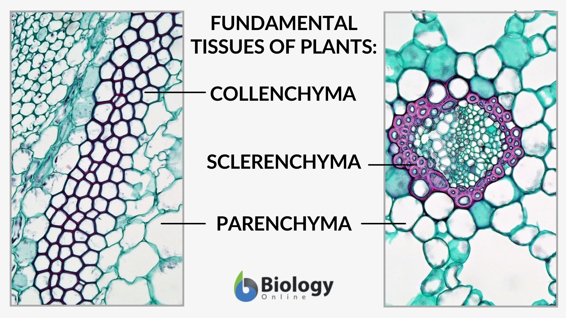
- Plant Cell Morphology: Plant cells have a distinct cell wall which gives plant cells a highly defined morphology. The shape varies from tissue to tissue. Epidermal cells are generally rectangular while cortex cells are isodiametric. Some sclerenchyma cells are stone-shaped while parenchyma cells range from nearly spherical to brick-like shape.

Figure 2: Different cell morphology in plants. Source: Maria Victoria Gonzaga of Biology Online, from the works (public domain) of Berkshire Community College Bioscience Library: cross-section of Cucurbita (common name: pumpkin or squash), magnification: 400x (left) and cross-section: Potamogeton leaf, magnification: 400x (right). &npbsp;
- Mammalian Cell Morphology: Mammalian cells are basically of 3 types:
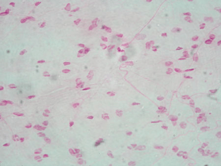
a) Epithelial-like cells: Rectangular shaped, kind of regular dimensions
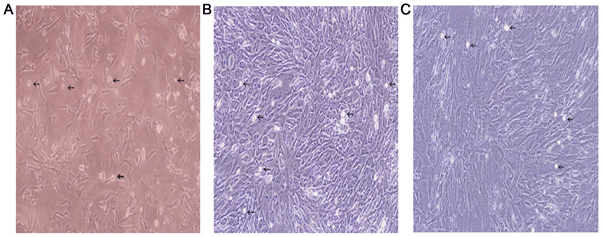
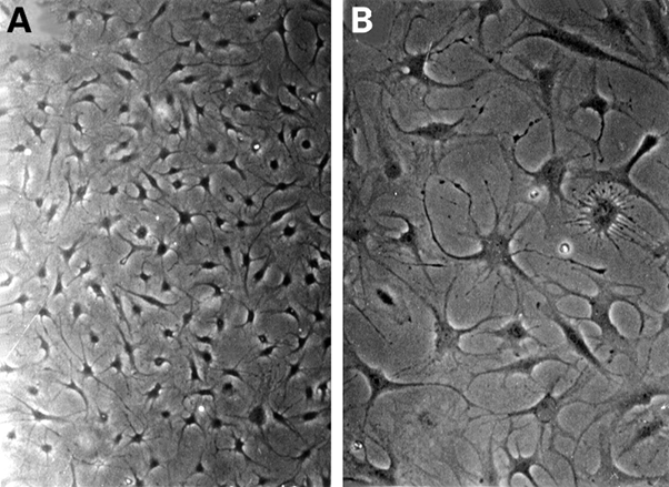
b) Fibroblast-like cells: Spindle shaped and flat, either bi- or multi-polar
{Both epithelial-like and fibroblast-like cells attach to a surface and grow!!}
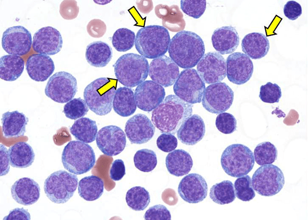
c) Lymphoblast-like cells: Spherical, round shaped, can grow in suspension (no need to attach to a surface!!)
Within these 3 major types, there are further sub-types. Like epithelial-like cells can be squamous, columnar, or cuboidal.
Let’s look at the table and figures below to learn about the different cell morphology examples in eukaryotes.
| Table 2: Different Cell Morphologies in Eukaryotes | |
|---|---|
| Cell Morphology | Examples of Eukaryotes |
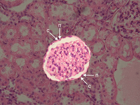
| Epithelial-like | Skin cells, cells in the lining of glomerular capsule in kidney, alveoli in lungs, endothelium in blood vessels’ cavities, pericardium in heart |
| Fibroblast-like | Areolar tissue cells, Fibroblast-like synoviocytes in joints, pannus lesions of patients with rheumatoid arthritis |
| Lymphoblast-like | Blood cells like RBCs, WBCs, cells of leukemia/lymphoma |
Interesting Fact!
Cell morphology is an exceptional field of Science that can help you in identifying when the cells of your body start behaving “abnormally”!!!
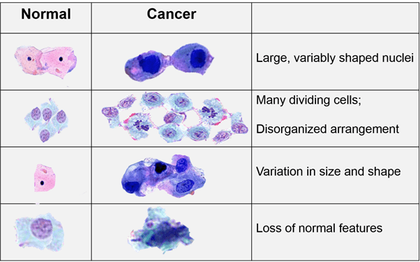
YES, just by closely observing a cell’s morphological characteristics like its shape, nucleus size, cytoplasmic content, invaginations, attachment to basal membrane, etc, we can tell if it is healthy or not. When a tumor cell grows in your body, it can be easily be distinguished because of the altered morphology- irregular shape, nucleus and nucleoli become bigger than normal, cytoplasm seems depleted, change in color of cytoplasm, detachment from the basal membrane in case of malignant tumors. These changes are indicative of cancerous growth! Also, for invasiveness and metastasizing, malignant tumors start producing some “lytic factors” which upon release, eat up the basal membrane and set the malignant cancerous cells “loose”.
Modern methods of in-vitro single cell-derived clones “MORPHO-PROFILING” can provide us with probable avenues and possibilities to identify cancerous and metastatic potentials of cells. This is how advancing Science paves way for precision medicines!!! Source: (Wu, 2020)
Try to answer the quiz below to check what you have learned so far about cell morphology.
Further Reading
References
- Chapter 4: Functional Anatomy Of Prokaryotic And Eukaryotic Cells. https://web.archive.org/web/20120923190032/http://www2.nemcc.edu/bkirk/Template%201/MICROCHAPTER4NOTES.htm
- Baron S. (1996) Medical Microbiology. 4th edition, Chapter 2- Structure https://www.ncbi.nlm.nih.gov/books/NBK8477/
- Zhao J., Ouyang Q., Hu Z., Huang Q., Wu J., Wang R., Yang M. (2016) A protocol for the culture and isolation of murine synovial fibroblasts. Biomedical reports, 5(2): 171-175 https://doi.org/10.3892/br.2016.708
- Xue C, Takahashi M, Hasunuma T. (1997) Characterisation of fibroblast-like cells in pannus lesions of patients with rheumatoid arthritis sharing properties of fibroblasts and chondrocytes. Annals of Rheumatic Diseases, 56:262-267.
- Wu P.H., Gilkes D. M., Phillip J. M., Narkar A., Cheng T. W.-T., Marchand J., Lee M.-H., Li R., Wirtz D. (2020) Single-cell morphology encodes metastatic potential. Sci. Adv. 6, 6938
©BiologyOnline.com. Content provided and moderated by Biology Online Editors.
