Cilium
n., plural: cilia
[ˈsɪl.i.əm/]
Definition: Microscopic hairlike projection in some eukaryotic cells
Table of Contents
Cilia Definition
Cilia are hair-like structures found on the surface of many types of cells, including some mammalian cells, especially those lining various tissues and organs. They are rudimentary and may be single or numerous. Cilia are important for movement. They also participate in mechanoreception. Ciliated organisms are those that have cilia. Their cilia are used for two main functions, they are to nourish or feed the organisms and to aid with movement.
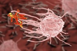
Cilia are typically studied as a part of cell biology, specifically, molecular cell biology in cell science. Cilia can be united into membranelles in short transverse rows or cirri in tufts. Cilia, which can beat in synchrony, transfer mammalian eggs through oviducts, generate water currents to carry food and oxygen past the gills of clams, transport food through the digestive systems of snails, circulate animal cerebral fluid, and remove detritus from mammals’ respiratory systems.
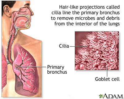
Cilia, in a modified form, cause jellyfish stinging devices to discharge and give rise to light-sensitive rods of the mammalian retina and odor-detecting units of olfactory neurons (sensory neurons). There are two types of cilia: motile and non-motile cilia, each with a subtype, for a total of four cell types. A cell will usually have one primary cilium or several motile cilia.
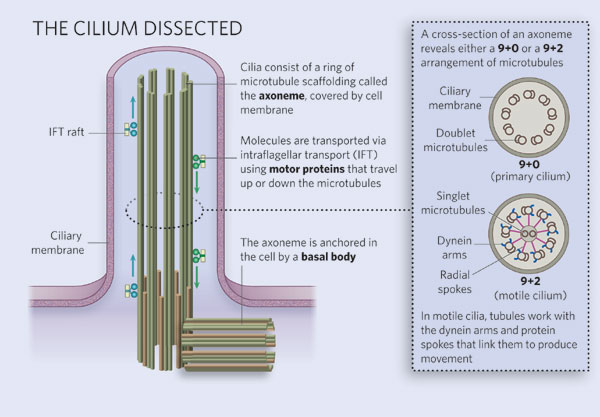
The cilium class is determined by the axoneme. The axoneme is the structure of the cilium core.
- When motile cilia function properly, they feature a 9+2 axoneme, which is composed of a core pair of single microtubules surrounded by nine outer microtubule doublets.
- The majority of non-motile cilia have a 9+0 axoneme lacking the central pair of microtubules. The accompanying components that permit motility (such as the outer and inner dynein arms and radial spokes) are also missing.
- Some motile cilia lack the center pair, while others do not, giving rise to the four varieties.
Watch this vid about cilia:
Biology definition:
Cilia are hair-like structures found on the surface of some cells. They are rudimentary in nature and may be single or numerous. Cilia are important for movement. They also participate in mechanoreception. Ciliated organisms are those that have cilia. Their cilia are used for feeding and movement. In botany, cilium (singular) refers to any of the hair forming a fringe along the margin or edge of a plant structure, e.g. of a leaf. Examples of organisms that have cilia are protozoans that use them for movement. In the human body, examples of tissue cells with cilia are the epithelia lining the lungs that sweep away fluids or particles.
Etymology: from Latin “cilium”, meaning “eyelid”
Structure
Cilia are membrane-bounded, centriole-derived cell surface projections that contain a microtubule cytoskeleton, the ciliary axoneme, and are enclosed by a ciliary membrane.
- Axonemes in multi-ciliated cells of mammalian epithelia are 9 + 2 and motile, with dynein arms.
- Cilia attach to cells through a basal body. The basal body is made up of nine triplets of microtubules. The triplets are created when the cilia’s doublets are connected by a different microtubule from the cell. Before approaching the basal body, the two central microtubules come to a stop.
- Motor proteins (dynein) are big, flexible molecules that enable cilia to move. For energy, the proteins hydrolyze ATP. When proteins are activated, they undergo conformational changes that allow them to move in complicated ways.
When a doublet is close by, the dynein molecules pull it up and reconnect further down the microtubules. The doublets can’t move very far and so are forced to make short-distance trips along each other since they are connected by cross-linking proteins. The cilium bends as a result of this movement.
Cilia are extremely small structures, measuring about 0.25 m in diameter and up to 20 m in length. They are seen in vast quantities on the cell surface when they are present. To create movement, the cilia work like oars, pounding back and forth.
The outer dynein arm (ODA) is a molecular complex that causes cilia/flagella to beat. Chlamydomonas ODA is made up of three heavy chains (HCs), two intermediate chains (ICs), and eleven light chains (LCs). The three-dimensional (3D) structure of the entire ODA complex has been studied, but the 3D configurations of the ICs and LCs are mainly unknown.
Basal body
The basal body, which refers to the mother centriole when it is linked with a cilium, is the cilium’s basis. Mammalian basal bodies comprise a barrel of nine triplet microtubules, subdistal appendages, and nine strut-like structures known as distal appendages that connect the basal body to the membrane at the cilium’s base. During axoneme growth, two of the basal body’s triplet microtubules expand to become doublet microtubules.
Ciliary rootlet
A cytoskeleton-like structure is a ciliary rootlet that develops from the basal body at the cilium’s proximal end. Rootlets are typically 80-100 nm in diameter and have cross striae spaced at regular intervals of 55-70 nm. Rootletin, a coiled-coil rootlet protein encoded by the CROCC gene, is a key component of the rootlet.
Transition zone
To attain its specific composition, the cilium’s proximal-most portion has a transition zone, also known as the ciliary gate, which regulates protein entry and exit from the cilium.
Y-shaped structures connect the ciliary membrane to the underlying axoneme at the transition zone.
Control of selective entrance into cilia may require a transition zone sieve-like function. Ciliaopathies, such as Joubert syndrome, are caused by inherited abnormalities in transition zone components. The structure and function of transition zones are conserved in a wide range of animals, including vertebrates (which have vertebrae cilia on their vertebrate cells), the roundworm, the fruit fly, and some specific algae.
Disruption of the transition zone in mammals lowers the ciliary abundance of membrane-associated ciliary proteins, such as those implicated in Hedgehog signal transduction, jeopardizing Hedgehog-dependent embryonic development of digit number and central nervous system patterning.
Types
Cilia are classified as either motile or non-motile. To generate fluid movement, motile cilia swing in a wave-like pattern. The surfaces of cells like epithelial cells of the upper respiratory tract and the reproductive tract have these cilia. Non-motile cilia cannot move and primarily serve as cellular antennas to regulate signaling pathways and maintain cell homeostasis.
Non-motile cilia
Primary cilia were identified in 1898 and are non-motile cilia. These structures were previously thought to be vestigial organelles. However, recent studies have revealed that primary cilia operate as a sensory cellular antenna that coordinates a huge number of cellular signaling pathways.
Modified non-motile cilia
Kinocilia located on inner ear hair cells are referred to as specialized primary cilia or modified non-motile cilia. They have the 9+2 axoneme of motile cilia but no inner dynein arms that allow movement. The outside dynein arms allow them to move passively in response to sound detection.
Motile cilia
These can be found in huge quantities on the cell’s surface. These are found in the respiratory epithelium of the respiratory system in humans. They do this by removing mucus and particles from the lungs.
Although the primary cilium was found in 1898, some believed it was a vestigial organelle with no vital role. Recent discoveries on its physiological roles in chemosensation, signal transduction, and cell growth control have highlighted its significance in cell function.
Its significance in human biology has been highlighted by the discovery of its role in ciliopathies, which are disorders caused by cilia dysgenesis or dysfunction, such as polycystic kidney disease, congenital heart disease, mitral valve prolapse, and retinal degeneration.
The primary cilium is now known to serve a critical role in the function of numerous human organs. Pancreatic beta cells’ primary cilia bend and regulate their function and energy metabolism. Islet dysfunction can result from cilia loss and type 2 diabetes.
» Modified motile cilia
Early embryonic development is characterized by motile cilia lacking the middle pair of singlets (9+0). They appear as nodal cilia on the primitive node’s nodal cells. In bilateral animals, nodal cells are responsible for left-right asymmetry.
Despite the absence of the central apparatus, there are dynein arms that allow the nodal cilia to spin. The movement causes a current flow of extraembryonic fluid across the nodal surface in a leftward direction, causing the developing embryo’s left-right asymmetry.
» Nodal cilia
A mono-cilium is a single cilium seen in nodal cells. They are present in the embryo’s very early development on the primitive node. The node has two sections with various forms of nodal cilia. The center node has motile cilia, and the nodal cilia on the node’s periphery are modified motile.
The leftward flow of extracellular fluid essential to start the left-right asymmetry is provided by the rotating motile cilia of the core cells.
Cilia versus flagella
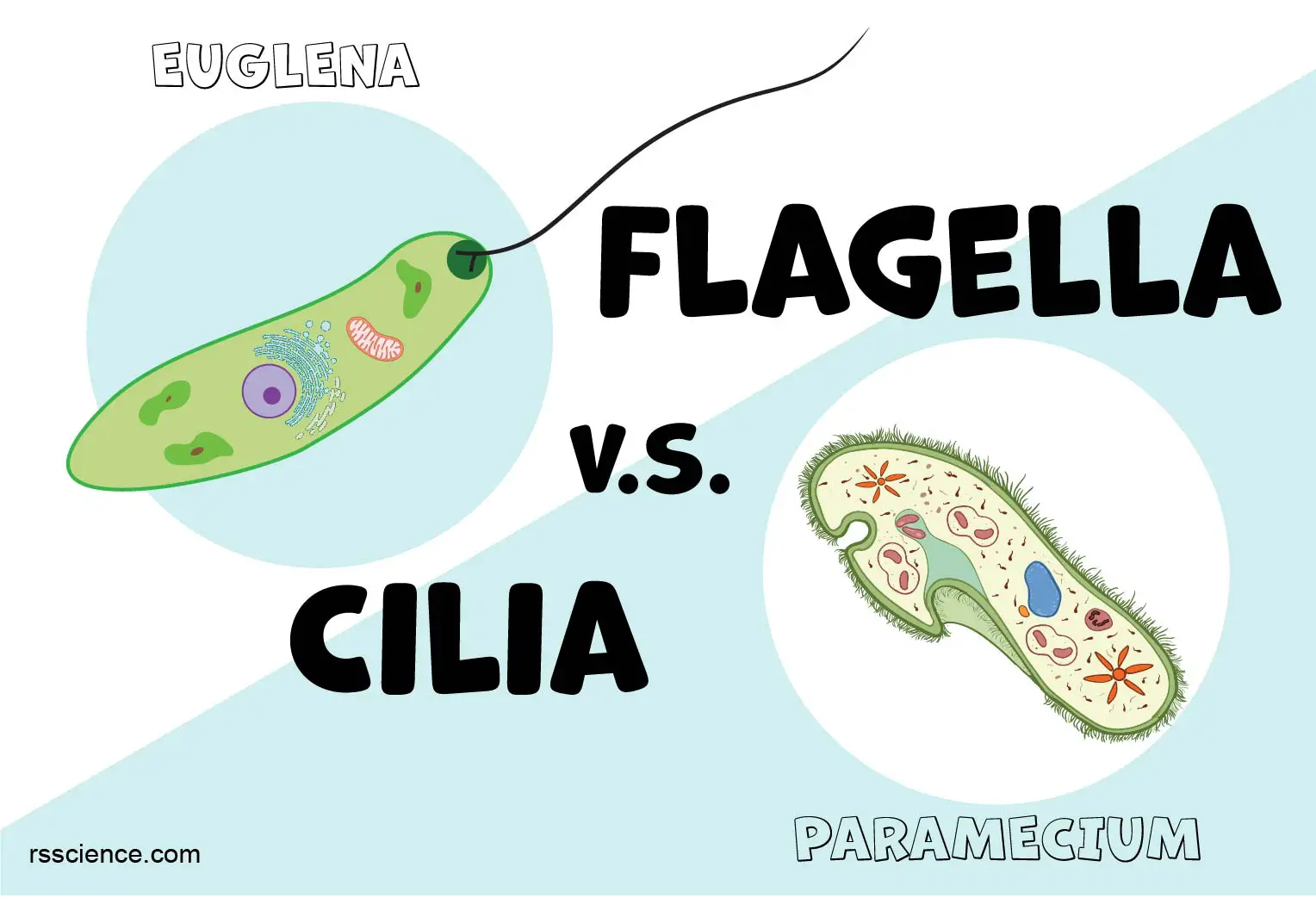
Flagella (singular: flagellum) as seen on sperm cells and numerous protozoans (flagellates) that allow them to float through liquids are sometimes referred to as motile cilia. Nevertheless, there are many references wherein they are used differently as there are some distinct differences between them. Eukaryotic flagella are “whip-like structures” and are often longer than conventional cilia and have a distinctive undulating motion.
Microorganisms
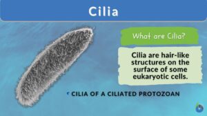
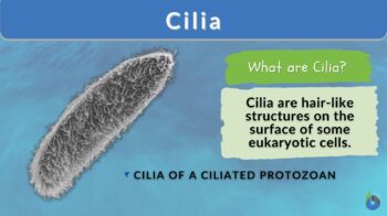
Ciliates are eukaryotic microorganisms that only have motile cilia and use them for movement or merely moving fluids across their surface. Paramecium, for example, has thousands of cilia that allow it to swim. These sensory motile cilia have been discovered. They are specifically called eukaryotic cilia.
Motility
Axonemal dyneins, which form sliding contacts between outer microtubules doublets, propel ciliary motility. Cilia motility intraflagellar transport proteins, another motor protein, convey materials into and out of the cilium. Ciliates are capable of intricate interactions with their surroundings. It follows that it is not unusual that occasionally sensory components (sensory cilia), such as insulin-like receptors, can be present on the cilium with other elements of signal transduction cascades.
Ciliogenesis
Ciliogenesis is the activity that occurs when either a cell’s antenna (primary cilia) or another part of the cell called the extracellular fluid mediation mechanism (motile cilium) is formed. During the cell cycle (cell division), it includes the building and disassembly of cilia.
Cilia are key cell organelles that participate in a variety of tasks such as cell signaling, signal processing, and controlling the flow of fluids such as mucus over and around cells. Because of the importance of these cell activities, ciliogenesis abnormalities can result in a variety of human diseases caused by non-functioning cilia. Ciliogenesis may also play a role in human left/right-handedness development.
Function
Cilia are essential for movement. This can include the cell itself moving, as well as other substances and things passing through the cell. Cilia are responsible for the overall movement of some organisms known as ciliates. Cilia, for example, cover the surface of the unicellular protist Paramecium and are responsible for mobility as well as eating. Cilia line the oral groove, which moves food into the organism’s “mouth” in addition to covering the outside of the creature.
Watch this vid of the Paramecium cilia movement:
Sensing the extracellular environment
In eukaryotes, some primary cilia on epithelial cells serve as cellular antennas, sensing the external environment through chemosensation, thermosensation, and mechanosensation. These cilia subsequently participate in the mediation of particular signaling cues, such as soluble substances in the extracellular environment, a secretory function in which a soluble protein is released to have an effect downstream of the fluid flow, and, if the cilia are motile, mediation of fluid flow.
Some epithelial cells have cilia on the apical surface, and these cells frequently form a sheet of polarized cells that forms a tube or tubule with cilia sticking out into the lumen. Cilia play a crucial part in maintaining the local cellular environment due to their sensory and signaling functions, which may explain why ciliary abnormalities are associated with such a wide spectrum of human diseases.
Axo-ciliary synapse
With the help of axo-ciliary synapses, serotonergic axons and primary cilia of CA1 pyramidal neurons (neuronal cells) communicate, changing the neuron’s epigenetic state in the nucleus – “a way to change what is being transcribed or made in the nucleus” – through this signaling that is distinct from that at the plasma membrane, which is also longer-term.
Clinical Significance
A variety of human disorders can be caused by ciliary defects. Cilia defects disrupt numerous important signaling pathways required for embryonic development and adult physiology, providing a reasonable explanation for the typically multi-symptom character of various ciliopathies.
Primary ciliary dyskinesia, Bardet-Biedl syndrome, polycystic kidney, and liver disease, nephronophthisis, Alström syndrome, Meckel-Gruber syndrome, Sensenbrenner syndrome, and some kinds of retinal degeneration are all examples of ciliopathies.
Ciliaopathies are chronic illnesses caused by genetic abnormalities that impair the proper functioning of cilia. They include Senior-Lken syndrome as well. Furthermore, a primary cilium deficiency in renal tubule cells can result in polycystic kidney disease (PKD). This affects urine flow.
The mutant gene products of another genetic condition known as Bardet-Biedl syndrome (BBS) are components of the basal body and cilia. Cilia cell defects have been linked to obesity and are frequently seen in type 2 diabetes. Several studies have already shown that ciliopathy models have poor glucose tolerance and reduced insulin secretion. Furthermore, the number and length of cilia were reduced in type 2 diabetes models.
Epithelial sodium channels (ENaCs) that are found along the length of cilia regulate the level of periciliary fluid. Mutations that reduce ENaC activity cause multisystem pseudohypoaldosteronism, which is linked to fertility issues. ENaC activity is increased in cystic fibrosis, which is caused by mutations in the chloride channel CFTR, resulting in a severe decrease in fluid level, which causes problems and infections in the respiratory cilia in the airways.
Cilia Disorders
Just like all body parts, if there are issues or disorders, it causes problems in the body. Here are some of the common human genetic disorders or ciliary defects that cause issues that were not mentioned previously in this article:
- Ciliopathies – It is a hereditary disease of cilia structures (basal bodies) or cilia function. Primary and motile cilia dysfunction or abnormalities are known to produce a variety of severe hereditary illnesses known as ciliopathies.
- Primary Ciliary Dyskinesia – It is an autosomal recessive condition characterized by abnormal cilia function. This disorder stops mucus from clearing from the lungs, ears, and sinuses.

Key Notes!
Cilia are organelles found in eukaryotic cells, especially in some animals and protozoans. They are classified into two types: motile cilia and non-motile cilia. Primary cilia are non-motile cilia that serve as sensory organelles. Cilia and flagella are structurally identical. Cilia are locomotory structures found in microorganisms such as Paramecium.
Take the Cilium – Biology Quiz!
References
- Cilia. (n.d.). Kenhub. Retrieved June 14, 2023, from https://www.kenhub.com/en/library/anatomy/cilia
- Cilia—Definition, Structure, Types & Function. (2020, October 7). BYJUS. https://byjus.com/biology/cilia/
- Cilium | Definition, Function, & Facts | Britannica. (n.d.). Retrieved June 14, 2023, from https://www.britannica.com/science/cilium
- Common Variants in Left/Right Asymmetry Genes and Pathways Are Associated with Relative Hand Skill—PMC. (n.d.). Retrieved June 14, 2023, from https://www.ncbi.nlm.nih.gov/pmc/articles/PMC3772043/
- CROCC – Rootletin—Homo sapiens (Human) | UniProtKB | UniProt. (n.d.). Retrieved June 14, 2023, from https://www.uniprot.org/uniprotkb/Q5TZA2/entry
- Editors, B. D. (2017, June 25). Cilium—Definition, Function and Structure. Biology Dictionary. https://biologydictionary.net/cilium/
- Fisch, C., & Dupuis-Williams, P. (2011). Ultrastructure of cilia and flagella—Back to the future! Biology of the Cell, 103(6), 249–270. https://doi.org/10.1042/BC20100139
- Garcia, G., Raleigh, D. R., & Reiter, J. F. (2018). How the Ciliary Membrane Is Organized Inside-Out to Communicate Outside-In. Current Biology: CB, 28(8), R421–R434. https://doi.org/10.1016/j.cub.2018.03.010
- Ishikawa, H., & Marshall, W. F. (2011). Ciliogenesis: Building the cell’s antenna. Nature Reviews. Molecular Cell Biology, 12(4), 222–234. https://doi.org/10.1038/nrm3085
- Johnson, K. A., & Rosenbaum, J. L. (1992). Polarity of flagellar assembly in Chlamydomonas. The Journal of Cell Biology, 119(6), 1605–1611. https://doi.org/10.1083/jcb.119.6.1605
- Lindemann, C. B., & Lesich, K. A. (2010). Flagellar and ciliary beating: The proven and the possible. Journal of Cell Science, 123(Pt 4), 519–528. https://doi.org/10.1242/jcs.051326
- Oda, T., Abe, T., Yanagisawa, H., & Kikkawa, M. (2016). Structure and function of outer dynein arm intermediate and light chain complex. Molecular Biology of the Cell, 27(7), 1051–1059. https://doi.org/10.1091/mbc.E15-10-0723
- Ringers, C., Olstad, E. W., & Jurisch-Yaksi, N. (2019). The role of motile cilia in the development and physiology of the nervous system. Philosophical Transactions of the Royal Society B: Biological Sciences, 375(1792), 20190156. https://doi.org/10.1098/rstb.2019.0156
- Rosenbaum, J. L., & Witman, G. B. (2002). Intraflagellar transport. Nature Reviews. Molecular Cell Biology, 3(11), 813–825. https://doi.org/10.1038/nrm952
- Satir, P., & Christensen, S. T. (2007). Overview of structure and function of mammalian cilia. Annual Review of Physiology, 69, 377–400. https://doi.org/10.1146/annurev.physiol.69.040705.141236
- Wheatley, D. N. (2021). Primary cilia: Turning points in establishing their ubiquity, sensory role and the pathological consequences of dysfunction. Journal of Cell Communication and Signaling, 15(3), 291–297. https://doi.org/10.1007/s12079-021-00615-5
©BiologyOnline.com. Content provided and moderated by Biology Online Editors.

