Colony Forming Unit
n., plural: colony forming units
/ˈkɑləni ˈfɔrmɪŋ ˈjunɪt/
Definition: Viable cell or group of cells forming a visible colony
Table of Contents
Colony Forming Unit Definition
A Colony Forming Unit (CFU) in microbiology and cellular biology refers to a measure of viable cells in a colony derived from a single progenitor cell. In microbiology, CFU is used to determine the number of viable bacterial cells in a sample per mL. Hence, it is usually used to indicate the degree of contamination in samples of water, vegetables, soil or fruits, or the magnitude of the infection in humans and animals. And by “viable” it means it includes only living cells (not the dead cells).
The term is fundamental in a microbiology or cell biology laboratory setting. A colony-forming unit represents a single viable cell or a cluster of cells that generates a visible colony under specific growth conditions. This unit serves as a fundamental measure of cell viability and growth potential that researchers use to quantify the presence or proliferation of microorganisms or cells within a given sample. This is important, especially in assessing or measuring the effectiveness of antimicrobial agents.
From One Cell to Billions – Colony Forming Units (by TheRubinLab):
A Colony Forming Unit represents a single viable cell or a group of cells capable of forming a visible colony under specific growth conditions. CFU in clinical microbiology, in particular, denotes either a single viable cell or a group of cells capable of forming a visible colony when plated under specific growth conditions. This concept is pivotal in various fields of biology, including microbiology, cellular biology, and pharmacology, as it serves as a fundamental unit to quantify viable microorganisms or cells. Etymology: The term “Colony Forming Unit” originates from the biological concept of colonies, referring to visible growth derived from a single or a group of cells, and the quantifiable unit of measurement. Acronym: CFU
Conceptualizing CFU
While it is difficult to trace the first use of the term, it is believed to have evolved within the microbiology and cellular biology science community in the late 19th and early 20th centuries, at about the time of the pioneering works of Louis Pasteur and Robert Koch. Louis Pasteur, in particular, laid the groundwork for understanding the microbial role in fermentation and the germ theory of disease. Both Pasteur and Koch helped shape the methods used in growing microbes in culture media like agar plates. In particular, Pasteur is credited for developing techniques for isolating and observing microbial colonies. Koche, in turn, refined techniques for isolating microbial colonies into individual colonies.
Microbial colony
The term “colony” in a microbiological context pertains to a visible cluster of microorganisms that originated from a single cell or group of the same cells on solid media. A colony in this sense can be likened to a colony in the macroscopic world, such as an ant colony or a bee colony. As for microorganisms, a bacterium cell, for instance, can replicate itself (by binary fission) and give rise to viable bacterial cells that shall be multiplying, too, and over time, they form an aggregate in a culture media that is visible to the naked eye. These “bacterial colonies” may display colony characteristics (color, texture, and morphology) that are distinct from other species.
Unit in the CFU
The term unit in the CFU may serve the following purposes:
- As a unit, it could mean it is an entity that is quantifiable.
- The “unit” in CFU serves as a tangible metric for assessing or identifying the efficacy of a treatment, for instance, or the viability of microorganisms in a given condition,
- The term “unit” represents a standardized measure that can be reproduced across different laboratory settings or studies,
The term “colony forming unit” could therefore be used to pertain to the standardized methods for quantifying microbial and cellular viability.
CFU Assay Applications
The importance of Colony Forming Unit assays can be observed in various domains. Here are some examples of its application in biological research and clinical practice:
- For studying microbial diversity
- For assessing microbial growth under certain conditions
- For evaluating the efficacy of antimicrobial agents (CFU analysis in drug discovery/ CFU and antimicrobial susceptibility testing)
- For diagnosing infections (identifying pathogens)
- For evaluating cell proliferation (Colony Forming Unit significance in quantifying cells)
- For assessing stem cell potency (CFU in stem cell research)
- For identifying and quantifying microbial communities in soil, water, or air samples
Steps In CFU Determination
- The process typically begins with the preparation of agar plates or other solid culture media.
- The sample containing the cells of interest is inoculated into the cell media.
- Inoculated plates are incubated under optimal conditions.
- Colonies form and are counted, typically using a colony counter or automated CFU counting methods.
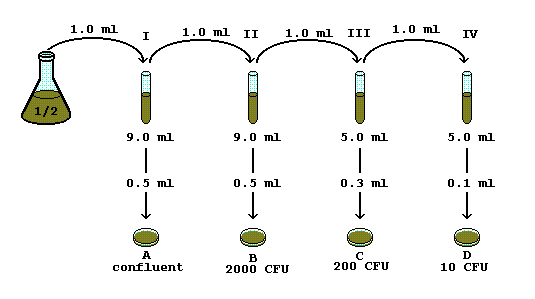
To ensure accurate CFU quantification, researchers often perform serial dilutions of the sample to obtain plates with an appropriate number of colonies for counting. Serial dilutions are essential for obtaining colonies on agar plates within a countable range. Serial dilutions involve the dilution of the sample in a series of tubes containing a known volume of diluent.

Calculating the CFU entails the use of this formula:
CFU/mL or CFU/g = Number of Colonies Counted ÷ (Volume Plated × Dilution Factor)
where,
Number of Colonies Counted: The total number of colonies counted on all plates
Volume Plated: The volume of the diluted sample plated onto the agar plate (in mL)
Dilution Factor: The dilution factor used during serial dilution (e.g., if you plated 0.1 mL of a 1:100 dilution, the dilution factor is 100)
CFU interpretation guidelines:
The value in CFU/mL (for liquid samples) or CFU/g (for solid samples) indicates the estimated concentration of viable cells in the original sample.
NOTE IT!
CFU assays may be influenced by certain factors. Hence, care must be taken when interpreting values. For instance, nutrient availability in culture media is one of the vital factors affecting the extent of microbial growth. Not enough nutrients available could mean diminished microbial growth. Environmental conditions like temperature, pH, and oxygen levels can also affect microbial growth. Take for instance the obligate anaerobes that are susceptible to the presence of oxygen. Other factors, such as inoculum size and incubation time, can alter microbial growth kinetics as well. These factors should be taken into account when conducting experiments employing CFU assays to ensure reliable and reproducible results that follow the standardization of CFU assays.
References
- Madigan, M. T., Bender, K. S., Buckley, D. H., Sattley, W. M., & Stahl, D. A. (2018). Brock Biology of Microorganisms. Pearson.
- Atlas, R. M. (2015). Principles of Microbiology. Jones & Bartlett Learning.
©BiologyOnline.com. Content provided and moderated by Biology Online Editors.


