Crown of head
n., [kɹaʊn ɔv ˈhɛd]
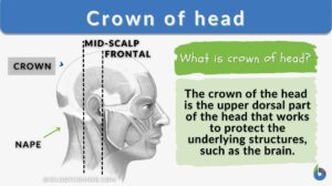
Definition: the area located at the upper dorsal (back) part of the head
Table of Contents
Crown of Head Definition
The crown of the head is the upper dorsal part (or area) of the head. Several creatures have diverse crown anatomy. It consists of the scalp that lies atop the skull, which in humans, consists of five layers. Moreover, a number of bone sutures are covered by the crown, which also houses blood vessels and trigeminal nerve branches.
Where is the Crown of Your Hair?
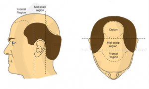
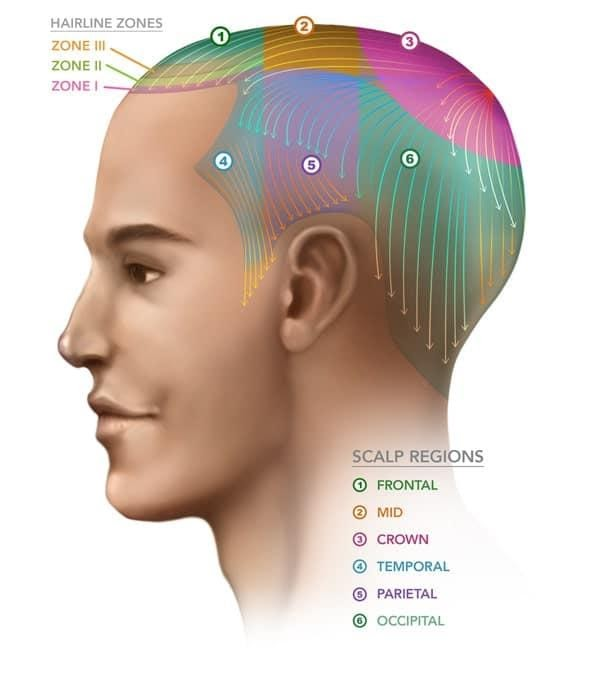
The dorsal upper portion of your skull is where the crown of your head is situated. It may also be referred to as the vertex on occasion. The crown functions to protect and support the tissues in your head, including your brain, just like other parts of your skull. It is situated near the sagittal suture, one of the joints that join the bones in your head.
Your skull’s sagittal suture runs down the middle from the front to the back. On this line, at its highest point, is where the actual crown is located. By placing a fingertip on the midline of your skull and starting to slide your fingers toward the back of your head, you may identify the crown of your head.
Biology definition:
The crown is the uppermost dorsal part of the head (or skull). It is also called corona capitis or, sometimes, “vertex”, which literally means the “highest point”.
Structure
One of the first things one should note is that there are many layers of the scalp located in the crown, which is at the top of the head. The different layers of the scalp are as follows (arranged from outer to innermost): Skin, Connective tissue, Aponeurosis, Loose connective tissue, and Pericranium.
Watch this vid about the layers of the scalp:
The crown encircles the skull’s bony layers. It varies from person to person and ranges in thickness from 0.16 to 0.28 inches or 4 to 7 millimeters. Over age, its thickness tends to rise.
The frontal bone and the parietal bone are divided below the crown by several fibrous junctions known as sutures. The sutures are a crucial component of growth and development because they allow the skull to enlarge as the brain gets bigger.
Human heads are symmetrically shaped because certain sutures between the frontal and parietal bones of the skull expand in particular directions. The frontal suture connects the parietal and frontal bones. The squamous part, the orbital part, and the nasal part are some of the components of the frontal bone.
Function
The crown’s primary purpose is to shield the brain from specific physical harm. The frontal and parietal bones, which make up the neurocranium’s crown and shield the frontal and parietal lobes of the brain, are present.
The meninges’ three membranes guarantee stability and shield these lobes from harm. For instance, the frontal lobe, situated behind the forehead, is intended to be protected by the thin meninges flexible sheets between the brain, spinal cord, and skull.
By serving as a cushion, the cerebrospinal fluid in the ventricles of the skull lessens the severity of the injury. Humans can conduct motor activities and movements by safeguarding the frontal lobe. The thick layers of the crown protect the parietal lobe of the neocortex, which has a strip that targets the sense of touch and enables the representation of space for action.
Injuries and Diseases
The brain is susceptible to a variety of illnesses and injuries that can affect the crown or human head. The number of illnesses and injuries affecting the human crown have different effects on the brain, which affects the person’s capacity for regular operation.
Many injuries and ailments have distinct causes, medical conditions, methods of diagnosis, and therapies. Some of the most common types of injuries or diseases are listed below. The most common type of disease or injury of the crown is one that is unexpected.
Hair Loss
Hair growth in humans starts at the skin and the hair comes out of hair follicles. When you start losing hair from your scalp or other areas of your body, this is called hair loss. This can be hair thinning, which affects the full head or body, or when persons lose hair in certain places altogether.
Some persons opt to use certain medications to regrow hair (depending on the hair growth cycle and its resting phase versus growing phase) in mild cases while in more severe cases may need a hair transplant (as seen in recent developmental biology).
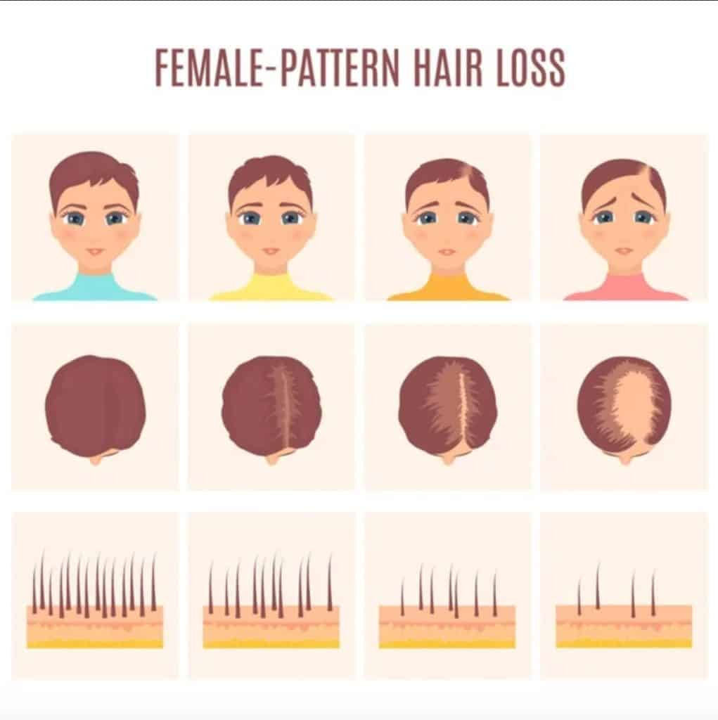
Another name for hair loss is alopecia. Androgenic alopecia and alopecia areata are two types of hair loss that usually affect the crown of your head. The most typical type of hair loss in both men and women is androgenic alopecia. Many persons who suffer from androgenic alopecia have a predisposition to the genetic condition.
Male pattern baldness and female pattern baldness have different hair loss patterns specifically, however androgenic alopecia is characterized by thinning hair at the crown of the head in both sexes.
This can be seen as a receding hairline (can be caused by high ponytails), loss at the crown, or bald patches. This may pose issues in extreme hot or cold weather as it exposes the crown area to the elements. Also, it’s probable that androgenic alopecia, which affects the crown of your head, will worsen the condition and put one more at risk for other health conditions.
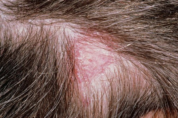
Ring Worm (Tinea capitis)
The scalp is one of the different parts of the body that ringworm, a fungus, can infect. Ringworm on the scalp is referred to as Tinea capitis. Children are more likely than adults to contract Tinea capitis.
Tinea capitis is transferred through close physical contact with an infected person or animal. Also, you can spread it by sharing private goods like caps, combs, and hairbrushes.
The infection first begins where the contact took place, but it has the potential to eventually spread across the whole scalp. The following are a few tinea capitis signs and symptoms:
- Scaly skin
- Itching
- hair loss
- hair that is brittle and readily breaks off, along with circular areas of skin that are red and irritated near the margins and progressively spread
This can sometimes cause permanent damage to the scalp if not treated properly and prevent new hairs from growing in certain areas.
Cerebrospinal fluid leak (CSF leak)
The cerebrospinal fluid leak, which entails the excessive loss of fluid from the meninges, is a common condition connected to the crown. The meninges membrane is primarily torn during a head, brain, or spinal injury, which results in the cerebrospinal fluid leak.
Intense headaches that are frequently centered on the crown are among the symptoms caused by excessive cerebrospinal fluid leakage. The patient’s ears and nose may leak fluid, which is a severe indication of this illness.
Examinations such as a computerized tomography scan, which produces an X-ray image of the crown and other areas of the skull, are used to diagnose the cerebrospinal fluid leak.
Meningioma
The cranial condition known as meningioma is characterized by the growth of tumors on the meninges, which surround the blood vessels and nerves close to the crown. Cells nearby have been known to divide quickly, which is one of the disorders’ causes.
Amnesia and epileptic seizures are among the indications and symptoms that meningioma patients experience. Arms and legs may become weak as a result of the frontal region of the brain being directly affected. Imaging studies, such as magnetic resonance imaging (MRI), which uses high-frequency radio waves and a strong magnetic field to measure the protons in the water, are used to make diagnoses.
Surgery is used to remove the tumor from the patient’s meninges as part of the best treatment, and the extent of the surgery is determined by the size and aggressiveness of the meningioma.
Fractures
According to the force of the impact on the skull, bone fractures to the crown of the head can be linear or depressed and vary in severity. A break in the skull occurs in a linear fracture, but a depressed fracture causes the fragments of the skull to scatter.
Most occurrences involving a vehicle, an assault, or a fall result in skull fractures. Penetrating skull fractures are more serious situations where an object, like a metal rod or bullet, entirely penetrates the skull.
Symptoms may include nausea, memory loss, concussion, bruising, and lethargy, depending on how severe the fracture is. Another symptom, such as bleeding, causes the skull’s contained cavity to build up pressure, which pushes the brain toward the brainstem opening and causes a coma.
Gorham Disease
The entire musculoskeletal system, particularly the crown of the skull, is affected by Gorham disease. Even though the chronic condition entails a steady loss of bone, the early signs and stages do not show severe discomfort.
Although the etiology of Gorham disease has not been identified, cells such as osteoclasts that are involved in the disintegration of weak and old bones are thought to be the key factor.
After a fracture to the crown of the skull, which results in aberrant deformities and problems with the neurological system in patients, the disease’s symptoms become obvious. Physical examinations, such as X-rays and magnetic resonance imaging, which detect abnormalities and a loss of bone mass (osteolysis), are used to make the diagnosis.
A variety of methods are used in the treatment of the disease to stop it from spreading from the patient’s skull to their spine or chest. Chemotherapy and surgery, as well as lifestyle changes such as consuming a diet of high protein, try to decrease the severity of the condition.
Embryonic Development
The formation of the crown of the head involves a series of embryonic events.
- Neural tube formation: referred to as neurulation, the neural tube forms from the ectoderm. The neural tube develops and eventually becomes the central nervous system, including the brain.
- Development of cranial neural crest cells: specialized cells from the neural tube called neural crest cells develop into parts of the head and face, including the crown region.
- Development of cranial mesenchyme and somitomeres: another group of cells, called mesenchymal cells, develop into cranial mesenchyme. Together with the somitomeres, these structures contribute to the formation of the head and skull.
- Development of dermis and epidermis: other cells of the ectoderm develop into the outer skin layer as well as facilitate the formation of hair follicles in the crown region. Genetic and environmental factors influence the positioning and patterning of hair follicles on the crown of the head.
- Formation of cranial bones: the mesenchymal cells differentiate into bone-forming cells, for the development of the cranial bones that make up the crown of the head.
- Vascularization: blood vessels develop to provide the developing structures with nutrients and oxygen.
Many of these processes occur simultaneously in a well-coordinated fashion to ensure the proper development of the crown of the head. Otherwise, improper development of the crown of the head could arise. Examples are as follows:
- Craniosynostosis, which refers to the premature fusion of the sutures or joints between the bones of the skull
- Anencephaly, a condition characterized by the failure of the neural tube to close as it should, thus, resulting in the absence of a major portion of the brain, skull, or scalp
- Microcephaly is a condition characterized by a head size that is relatively smaller than the average size expected of an individual’s age. The brain did not develop or grow properly leading to a smaller skull due to a variety of factors, such as genetic mutations or exposure to certain pathogens, such as the Zika virus.
Evolution
Compared to monkeys, the human species underwent macroevolution, which produced modifications such as an expansion in the bone and muscle structures that support the crown of the skull.
Note it! Baby’s crown
Babies have very soft crowns although the crown of the head is typically hard in an adult. Fontanels are two significant soft patches that are present at birth on the crown of the head. These soft areas are the gaps between the skull’s bones when bone growth hasn’t finished. As a result, the skull can be shaped during birth.
Compared to archaic human species, modern human species have a more angular skull base and a rounder cranial vault. The temporal lobes of the modern human species are located beneath the base of the skull, indicating an increase in the size of the human brain and skull.
Archaic and modern human species share the same architecture of the sagittal vault, the region that connects the two parietal bones to form the structure of the crown. The modifications of the crown are largely influenced by the cartilage that is lodged within the skull.
The cranial cartilage played a crucial role in protecting the central nervous system, but over time, a process called endochondral ossification caused the cartilage to start molding the crown. To create the skull’s bone structure, growing cartilage is replaced by bone during this process.
Take the Quiz!
References
- Akhtari, M., Bryant, H. C., Mamelak, A. N., Flynn, E. R., Heller, L., Shih, J. J., Mandelkern, M., Matlachov, A., Ranken, D. M., Best, E. D., DiMauro, M. A., Lee, R. R., & Sutherling, W. W. (2002). Conductivities of three-layer live human skull. Brain Topography, 14(3), 151–167. https://doi.org/10.1023/a:1014590923185
- Berger, A. (2002). Magnetic resonance imaging. BMJ (Clinical Research Ed.), 324(7328), 35. https://doi.org/10.1136/bmj.324.7328.35
- Brain Hemorrhage Symptoms, Treatment, Types, Recovery Time. (n.d.). Retrieved March 26, 2023, from https://www.medicinenet.com/brain_hemorrhage/article.htm
Cerebrospinal Fluid (CSF) Leak—Symptoms and Causes. (n.d.). Retrieved March 26, 2023, from https://www.pennmedicine.org/for-patients-and-visitors/patient-information/conditions-treated-a-to-z/cerebrospinal-fluid-leak - Crown of Head: Conditions, Injuries, and More. (2020, November 18). Healthline. https://www.healthline.com/health/crown-of-head
- Graves, G. R. (1990). Function of Crest Displays in Royal Flycatchers (Onychorhynchus). The Condor, 92(2), 522–524. https://doi.org/10.2307/1368252
- Hendricks, B. K., Patel, A. J., Hartman, J., Seifert, M. F., & Cohen-Gadol, A. (2018). Operative Anatomy of the Human Skull: A Virtual Reality Expedition. Operative Neurosurgery (Hagerstown, Md.), 15(4), 368–377. https://doi.org/10.1093/ons/opy166
- Kaucka, M., & Adameyko, I. (2019). Evolution and development of the cartilaginous skull: From a lancelet towards a human face. Seminars in Cell & Developmental Biology, 91, 2–12. https://doi.org/10.1016/j.semcdb.2017.12.007
- Meningioma—Symptoms and Causes. (n.d.). Retrieved March 26, 2023, from https://www.pennmedicine.org/for-patients-and-visitors/patient-information/conditions-treated-a-to-z/meningioma
- Ross, J. L., Schinella, R., & Shenkman, L. (1978). Massive osteolysis. An unusual cause of bone destruction. The American Journal of Medicine, 65(2), 367–372. https://doi.org/10.1016/0002-9343(78)90834-3
- Sakka, L., Coll, G., & Chazal, J. (2011). Anatomy and physiology of cerebrospinal fluid. European Annals of Otorhinolaryngology, Head and Neck Diseases, 128(6), 309–316. https://doi.org/10.1016/j.anorl.2011.03.002
- The evolution of the human head | WorldCat.org. (n.d.). Retrieved March 26, 2023, from https://www.worldcat.org/title/726740647
- Voo, K., Kumaresan, S., Pintar, F. A., Yoganandan, N., & Sances, A. (1996). Finite-element models of the human head. Medical & Biological Engineering & Computing, 34(5), 375–381. https://doi.org/10.1007/BF02520009
- What is a Skull Fracture? | UC Health | Symptoms & Causes. (n.d.). Retrieved March 26, 2023, from https://www.uchealth.com/en/conditions/skull-fractures
©BiologyOnline.com. Content provided and moderated by Biology Online Editors.

