Ectoplasm
n., plural: ectoplasms
[ˈɛktəˌplæzəm]
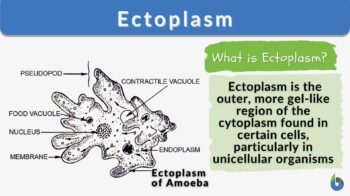
Definition: The outer, more liquid and transparent region of the cytoplasm found in certain cells
Table of Contents
Definition Of Ectoplasm
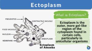
The peripheral part of the cytoplasm which is clear, gel-like, rigid, and agranular part is known as ectoplasm. Ectoplasm comes from the Greek word, ‘ektos’ (meaning “outside”) and ‘plasma’ (meaning substance having a form). The term “ectoplasm” was first used in the scientific context. The German biologist and philosopher Ernst Haeckel is credited as the person to have coined the term in the late 19th century when he refer to the clear, outer region of the cytoplasm in unicellular organisms such as amoebas (in contrast to the inner region he referred to as endoplasm). In amoeba, ectoplasm is the driving force for the formation of pseudopodia. Contraction of ectoplasm causes cytoplasmic streaming causing pseudopodia formation and the movement of amoeba.
Later on, in the early 20th century, the term “ectoplasm” has been adopted by spiritualists in the context of spiritualism and psychical research. In the spirit world, ‘ectoplasm’ refers to the ‘ectoplasmic manifestation’ in the form of spiritual energy that is claimed to have emanated from mediums, particularly during séances or other spiritualistic practices. This manifestation of spiritual energy is described as a semi-transparent, gel-like substance, reminiscent of the ‘ectoplasm’ in biological context. These spiritual phenomena, though, in the paranormal context are regarded as pseudoscientific and lack empirical evidence.
Let us now focus on the definition of ectoplasm in the context of biology.
Ectoplasm in Biology
Biology definition:
An ectoplasm is the outer, non-granulated, clear portion of the cytoplasm of a cell of certain species. In amoeba, for instance, the cytoplasm is divided into endoplasm and ectoplasm. The inner dense and often granulated part of the cytoplasm is the endoplasm. The clear outer portion of the cytoplasm is the ectoplasm. While the endoplasm is adjacent to the nuclear envelope the ectoplasm lies immediately to the plasma membrane. Thus, the endoplasm houses the endomembrane system, which makes the endoplasm metabolically active. The ectoplasm, in turn, contains large numbers of actin filaments, and as such is associated with providing an elastic support for the cell membrane.
Synonyms: exoplasm
Compare: endoplasm
In biological science, ectoplasm is the aggranulated, gel-like, clear part of the cytoplasm which is adjacent to the plasma membrane. Ectoplasm forms the peripheral zone of the cytoplasm, which adheres to the plasma membrane surface.
The concept of ectoplasm is associated with eukaryotic cells. In a eukaryotic cell, all the cellular material enclosed in the cell membrane, except the nucleus constitutes the cytoplasm. The contents of the nucleus present inside the nucleus membrane are referred to as nucleoplasm. Cytoplasm is made up of multiple organelles, gel-sol-like material known as cytosol, and cytoplasmic inclusions.
- In some animal cells, the ectoplasm and the endoplasm form the cytoplasmic constitution. Endoplasm is the granulated part of the cytoplasm which is separated from the nucleus by the nuclear envelope. Thus, broadly speaking ectoplasm is the outer part of the cytoplasm while the endoplasm constitutes the inner part of the cytoplasm.
- Protozoans are one-cell or single eukaryotes wherein the cell is similar to an animal cell and possesses cell organelles (like endoplasmic reticulum, ribosomes, mitochondria, Golgi bodies, and nuclei). The cytoplasm/protoplasm in protozoans is distinctly divided into two zones: ectoplasm and endoplasm. Ameoba is a widely studied protozoan having ectoplasm and endoplasm. In amoeba, the ectoplasm and endoplasm primarily function in the locomotory action of the pseudopodia in amoeba. The ectoplasm is the outer cytoplasmic layer which is clear while the endoplasm is the inner cytoplasmic layer which is rich in granules.
- In amoeba, the ectoplasm is a semi-solid gelatinous substance (also known as the plasma gel) while the endoplasm is relatively less viscous (also known as plasmasol). Ectoplasm is present below the plasmalemma. Characteristically, ectoplasm is aggranulated, clear, and watery and is present adjacent to the plasma membrane. (Figure 1)
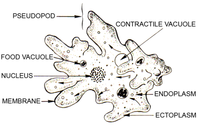
Ectoplasm vs. Endoplasm
The ectoplasm is a high surface tension layer. Also, ectoplasm is rich in actin filaments that provide support to the elasticity of the cell membrane. The cross-linking of actomyosin is responsible for the viscous nature of endoplasm. Being a gel-like structure, ectoplasm helps to protect the amoeba cell. On the other hand, endoplasm is dense, granulated, and found between nuclear plasm and ectoplasm.
The endoplasm is rich in granules, inorganic ions, enzymes, nucleic acid, water, carbohydrates, amino acids, and lipids. The endoplasm is highly metabolically active and houses most of the cell organelles. All the synthesis of essential elements for cell survival is synthesized as well as metabolized in the endoplasm.
Let us understand the difference between endoplasm and ectoplasm.
Table 1: Ectoplasm vs. Endoplasm | |
|---|---|
| Ectoplasm | Endoplasm |
| Outer, aggranulated part of the cytoplasm | Inner, granulated part of the cytoplasm |
| Transparent (clear ectoplasm), gel-like | Less viscous and non-transparent |
| Granules are absent | Granules are present along with cell organelles, enzymes, lipids, carbohydrates, proteins, and inorganic ions |
| Not dense | Highly dense |
| A very small portion of the cytoplasm and cell constitutes ectoplasm | A substantial bulk of the cytoplasm is the endoplasm |
| It is found adjacent to the cell membrane | It is adjacent to the nucleus and far from the cell membrane |
| Not many cellular processes occur in the ectoplasm | The major site for all the cellular and metabolic processes of a cell |
| Ectoplasm is more gel-like fluid wherein the gel-like consistency results in alveoli-like formations | The endoplasm is a kind of emulsion with granules and organelles suspended in it. |
| Ectoplasm is semi-permeable in nature | Endoplasm is hypertonic |
Data Source: Dr. Amita Joshi of Biology Online
Watch this vid about ectoplasm:
Role Of Endoplasm In Amoeba
The movement or locomotion in amoeba is through the formation of pseudopodia. Ectoplasm and endoplasm both work in combination for the movement in amoeba. In amoeba, the ectoplasm is rigid, contractile, thin, and clear part of the cytoplasm. The ectoplasm is responsible for the formation of pseudopodia. Further, ectoplasm also determines the change in the direction of movement by pseudopodium.
Change in the pH, i.e., acidity or alkalinity of the water, induces the variation in the ectoplasm which changes the direction of the ectoplasm. Even the slightest change in the pH of the water results in a change in the direction of the movement by pseudopodium. Osmotic pressure and ionic changes induce sol-gel conversion events which result in the event of contraction and relaxation that results in the locomotory action in the protoplasm of amoeba. (Sol-gel or Gel-sol is a known form of conversion of state of matter)
When an amoeba moves, the gelatinous ectoplasm extends, thereafter the plasma sol or the fluid-like endoplasm starts to flow towards the gelatinous ectoplasm. Later, the fluid-like endoplasm congeals towards the end of the pseudopodia. This results in the formation of the extended appendage. Simultaneously, at the opposite end of the amoeba cell, the plasma gel or the ectoplasm undergoes gel-sol conversion and forces the movement of the protoplasm towards the pseudopodia.
In the endoplasm, the breaking of the actin and myosin complex results in a more fluid-like ectoplasm that forces the endoplasm towards the pseudopodia. This causes relaxation movement in the amoeba. Later, the actin and myosin complex are reformed resulting in contractile movement in the amoeba. Such repeated relaxation-contraction movement due to gel-sol conversion in the protoplasm results in the movement of the amoeba cell. (Figure 2)
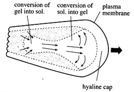
During the entire locomotion process, the endoplasm undergoes continuous gelation at the anterior tip of the pseudopod which results in the extension of the pseudopod while the ectoplasm undergoes sol formation at the posterior end of the pseudopod.
The ectoplasm at the posterior end of the pseudopodia pushes and squeezes the endoplasm to move in the direction of the pseudopodia. The ectoplasm at the posterior end of the pseudopodia consequently acts as the control center for the movement of the amoeba. Thus, different consistency of the endoplasm and ectoplasm help in the locomotion of the amoeba. Consequently, there is continuous conversion of ectoplasm to endoplasm and vice versa.
There is cyclic conversion of ectoplasm to endoplasm at the posterior end of the pseudopodia while endoplasm gets converted to ectoplasm at the anterior end or the tip of the pseudopodia. This produces a cyclic movement inside the amoeba which eventually results in the movement of the amoeba.
During the movement of the amoeba, the contraction of the ectoplasm causes the generation of internal hydrostatic pressure which results in the pressure flow of the endoplasm or the streaming of the endoplasm. Thus, contraction of the ectoplasm is the driving force for the cytoplasmic streaming that results in the movement of the amoeba.
Further, ectoplasm forms an ectoplasmic tube, when amoeba comes in contact with food. Later the ectoplasmic tube is converted to a vacuole that engulfs the food material inside it.
NOTE IT!
- The ectoplasmic contraction is unidirectional and the maximum contractile force that has been measured is 180 g.cm-2.
- Another important fact to remember is that like amoeba, in the human body, macrophages also exhibit locomotion using pseudopodia. Similar to amoeba, ectoplasm is the driving force for the movement of macrophages.
What Does The Ectoplasm Consist Of?
Ectoplasm is a clear, gel-like part of the cytoplasm present below the plasmalemma. Ectoplasm is aggranulated, i.e., devoid of granules. Ectoplasm has multiple longitudinal ridges. Ectoplasm encloses endoplasm from all sides.
Apart from other components, ectoplasm is rich in actin filaments and intermediate filaments. The presence of fine threads /filaments of actin provides support to the plasma membrane and also helps in maintaining elasticity. The actin filaments combine with the heavy meromyosin (HMM). These actomyosin complexes are prominently seen in the ectoplasmic tubes. Also, some of the actin-HMM complexes are attached to the plasmalemma. Thus, the actomyosin microfilaments are prominently found in the ectoplasmic tubes that usually terminate at the invaginated membrane surface.
Both G-actin and F-actin are present in the ectoplasm that undergoes G-F transitions. G-actin is the actin having a globular structure while the F-actin is the actin having a filamentous structure. G-actin is usually formed under low ionic concentrations while under the action of ATP molecules and metallic cations, the double helical structure of F-actin forms. With the help of ATP molecules, the G-actin gets converted to F-actin.
What Is The Function Of Ectoplasm?
Ectoplasm is the rigid, gel-like, clear layer of cytoplasm present underneath the plasmalemma. It encloses endoplasm from all sides. Endoplasm forms the bulk of the cytoplasm containing all the cell organelles. Thus, ectoplasm act as a protective barrier to the endoplasm and the cell organelles.
- Contraction of the ectoplasm is the primary driving force for the formation of pseudopodia in amoeba. Thus, ectoplasm is responsible for the locomotion/ movement in the amoeba.
- As we know, ectoplasm is relatively rigid and thus, it helps to maintain the structure of the cell.
- Ectoplasm is rich in actin fibers/filaments. The web of actomyosin microfilaments is present in the endoplasm and some of its part terminates in the plasmalemma. This helps in providing structural support and elasticity to the cell.
- The actin fibers present in the ectoplasm are also involved in the formation of spindle fibers during cell reproduction (mitosis as well as meiosis).
Review Questions
Based on the above discussion, let us answer some frequently asked questions.
- What is plasmagel?
- Endoplasm
- Ectoplasm
- Cytoplasm
- Epithelial cells
- Answer: B. Plasmagel or the ectoplasm is the gel-like, clear, rigid part of the cytoplasm present adjacent to the cell membrane.
- Part of cytosol that is present beneath the plasmalemma
- Hyalosome
- Nucleosome
- Plasmagel
- Plasmasol
- Answer: C. Plasmagel or endoplasm is present beneath the plasmalemma in amoeba
- Conversion of plasmsol to plasmagel is a
- Reversible, biochemical process
- Irreversible, physiological process
- Reversible, physical process
- Reversible, physical, and biochemical process
- Answer: D. Interconversion of plasmasol to plasmagel and vice versa is a physical conversion of gel state to sol state which is induced by biochemical changes.
- The movement of pseudopodia in macrophages is caused by
- Contraction of microfilaments in ectoplasm
- Turgor pressure changes
- Vacuoles are the driving force
- Oxygen availability
- Answer: A. Movement in macrophages is similar to amoeba and contraction of actin microfilaments in the ectoplasm is the driving force for the pseudopodia formation and movement thereof.
Take the Ectoplasm – Biology Quiz!
References
- Callan-Jones, A. (2022). Self-organization in amoeboid motility. Frontiers in Cell and Developmental Biology, 10, 1000071.
- Mast, S. O. (1926). Structure, movement, locomotion, and stimulation in amoeba. Journal of Morphology, 41(2), 347-425.
- Nascimento-Silva, G., Hardoim, C. C. P., & Custódio, M. R. (2022). Addressing the neglected associations between sponges and protists: The Porifera microeukaryome. Ficha Catalográfica, 41.
- Taylor, D. L., Rhodes, J. A., & Hammond, S. A. (1976). The contractile basis of ameboid movement. II. Structure and contractility of motile extracts and plasmalemma-ectoplasm ghosts. The Journal of cell biology, 70(1), 123–143. https://doi.org/10.1083/jcb.70.1.123
- Guyton, A. C. & Hall, J. E. Textbook of Medical Physiology, Eleventh Edition. Saunders.
©BiologyOnline.com. Content provided and moderated by Biology Online Editors.

