Elastic cartilage
n., plural: elastic cartilages
[əˈlæstɪk ˈkɑː.tɪl.ɪd͡ʒ]
Definition: a type of cartilage characterized by being rich in elastic fibers
Table of Contents
The cartilage is a connective tissue characterized by having an extracellular matrix that is abundant in chondroitin sulfate and chondrocytes (the cellular component). The cartilage has three main types: (1) elastic cartilage, (2) hyaline cartilage, and (3) fibrocartilage.
They differ mainly from the fibers that are present. In this article, we will focus on elastic cartilage. Let us understand what cartilage is, what’s it made of, its function, and its location in the body.
Elastic Cartilage Definition
Elastic cartilage tissue or yellow cartilage is the connective tissue that is found in body organs that do not bear body load, for example, the nose, ear, eustachian tube, and epiglottis. Elastic cartilage is a type of dense connective tissue. Elastic cartilage connective tissue functions to impart strength and elasticity to the organ. One of the major elastic cartilage characteristics is that it is rich in elastic fibers.
What does elastic cartilage look like?
The elastic cartilage is also known as yellow cartilage due to its yellowish appearance. The elastic fibers are responsible for the yellowish appearance of the tissue and also provide it with high elasticity. External ears, larynx, eustachian tube, and epiglottis have elastic cartilage.
Apart from elastic cartilage, the body has two other types of cartilage tissue; hyaline cartilage and fibrous cartilage are the other two types of cartilage. Elastic cartilage is similar in structure to hyaline cartilage, however, elastic cartilage has a dense network of elastic fibers. These elastic fibers are responsible for the elasticity and yellowish color of the elastic cartilage. The elastic fibers in elastic cartilage provide the ability for the tissue to return to its original form, post-stretching. Also, elastic cartilage does not have blood and nerve supply.
Biology definition:
Elastic cartilage is a type of connective tissue that is found in the body organs that do not bear the load. The cells are surrounded by a territorial capsular matrix and the interterritorial matrix containing elastic fiber networks, in addition to the collagen fibers and ground substance. Histologically, it resembles hyaline cartilage. Nevertheless, it can be distinguished from hyaline cartilage in being rich in elastic fibers. It is found in the external ear, nose, eustachian tube, and epiglottis. The primary function of elastic connective tissue is to provide strength and resilience to the organ.
Synonym: yellow cartilage
Compare: fibrocartilage; hyaline cartilage
Elastic Cartilage Structure
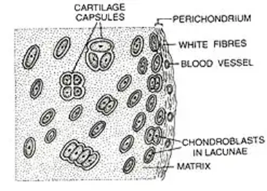
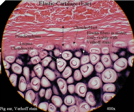
Structurally, elastic cartilage resembles a lot of hyaline cartilage, however, elastic cartilage has predominantly elastic fibers in its matrix and perichondrium. The chondrocytes are housed individually or in multiples, in spaces known as lacunae within the elastic fiber network.
In general, the presence of elastic fibers imparts elasticity to the elastic tissue like ear pinnae, lungs, larynx, etc.
What is cartilage made of?
Elastic cartilage histology reveals that it has three basic parts:
- specialized cells
- collagen and other fibers
- extracellular matrix

Note it!
Elastic cartilage is yellow in appearance. Hyaline and fibrous cartilages are white in appearance.
The chondrocytes and chondroblasts are the specialized cartilaginous cells. As compared to hyaline cartilage the number of chondrocytes is more in elastic cartilage.
The extracellular matrix and the chondrocytes are enclosed within an outer fibrous membrane known as perichondrium. The perichondrium, a connective tissue, surrounds cartilage. The perichondrium has two layers — inner perichondrium and outer perichondrium.
The chondroblasts secrete the extracellular matrix. The chondroblasts are the mesenchymal progenitor cells that will eventually form chondrocytes and are found near the outer layer of the perichondrium. (Figure 2)
During the process of secreting the extracellular matrix, the chondroblasts cells mature and are entrapped within the lacunae. The elastic cartilage matrix is primarily composed of type II collagen fibers, elastic fibers, water, and proteoglycans (aggrecans).
Aggrecans are the specific glucosaminoglycan that is found in cartilages only. They form an aggregate with hyaluronic acid. Interestingly, the negative charge on aggrecans is responsible for its water uptake capacity, imparting a cushion-like structure to the cartilage.
The collagen fiber provides strength to the cartilage while the elastic fibers in the elastic cartilage provide elasticity to the cartilage.

Watch this vid to see what elastic cartilage looks like under the microscope:
Note it!
Verhoeff van Gieson stain, Orcein, Aldehyde fuchsin, and Weigert’s elastic are some of the stains used to differentiate between hyaline cartilage and elastic cartilage. Verhoeff van Gieson stains the elastic fiber in black color.
Elastic Cartilage Functions
In Outer Ear
Elastic cartilage forms the external ear or the ear pinnae. Elastic cartilage provides the required flexibility to the external ear. The external ear is devoid of any nerve supply and is avascular. This ensures that the ear can fold during various day-to-day activities without any pain. Also, due to the elasticity, the external ear can withstand external trauma and funnel the sound vibration to the middle ear without any modification. Thus, a cartilage ear is required for normal functioning of the body.
Thickening of the ear cartilage may appear as a blue, gray, or black spot on ear cartilage and is known as ochronosis.
In Epiglottis and Larynx
The epiglottis is a leaf-shaped flap-like structure that is present over the laryngeal inlet. The elasticity and the flexibility of the epiglottis allow it to carry out two important functions of life.
One of the epiglottis functions is to cover the laryngeal inlet while swallowing the food. This prevents food or liquid from entering the lungs. The other function that epiglottis is involved in is breathing. Thus, the epiglottis functions like a valve that helps in normal breathing and prevents choking due to entry of food/liquid in the air passage or trachea.
The larynx is the voice box of the human body. The corniculate cartilage and cuneiform cartilage of the larynx are elastic cartilages. Together these cartilages work to control and modulate the vibrations generated in the voice box thereby affecting the voice quality and pitch.
In Eustachian Tube
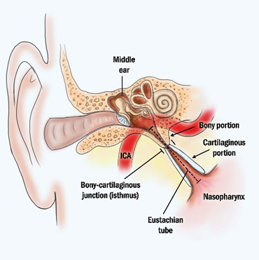
The eustachian tube or tuba auditive or pharyngotympanic tube is the connecting tube between the middle ear and the back of the throat or the nasopharynx. (Figure 3) The eustachian tube functions to drain out the mucus from the ear and balance the air pressure between the external environment and the middle ear.
Normally, the eustachian tube is closed thus preventing the infection to enter and invade the ears and prevent any unwanted imbalance due to changes in the atmospheric pressure. However, once in a while the eustachian tube opens up for physiological activities like yawning, in order to balance out the atmospheric pressure with that of the internal ear pressure.
The eustachian tube is composed of elastic cartilage and thus it has the strength, to retain its structure, along with the required flexibility, that allows the eustachian tube to come back to its original shape after balancing out the air pressure.

Where Is Elastic Cartilage Found?
Elastic cartilage location: Elastic cartilage is found in the external ear or ear pinnae, eustachian tube, larynx, and epiglottis. The elastic cartilage in these organs provides strength and flexibility which is necessary and critical for the normal physiological function of the body. Hope that answers common questions like what type of cartilage is found in the ear… what type of cartilage is found in the larynx… what is the external ear cartilage type… etc.
The Extracellular Matrix of Elastic Cartilage
The primary composition of the extracellular matrix of the elastic cartilage includes proteoglycan aggrecan, elastin fibers, glycoproteins, and collagen type II, IX, X & XI.
Elastin proteins are the primary protein present in the elastic cartilage. The presence of proteoglycan aggrecan is negatively charged and hence it attracts water molecules, thus giving a gel-like structure to the elastic cartilage. This gel-like structure imparts the property to withstand compressional forces.
The elastin fibers are present in the matrix as well as the perichondrium of the elastic cartilage provides flexibility to the elastic cartilage. Many elastic fibers are intermingled with type II collagen fibers.
Answer the quiz below to check what you have learned so far about elastic cartilage.
References
- Chang, L.R, Marston, G., Martin, A. (2021). Anatomy, Cartilage. In: StatPearls [Internet]. Treasure Island (FL): StatPearls Publishing; 2022 Jan-. Available from: https://www.ncbi.nlm.nih.gov/books/NBK532964/
- Cox, R. W., & Peacock, M. A. (1979). The growth of elastic cartilage. Journal of anatomy, 128(Pt 1), 207–213.
- Krishnan, Y., & Grodzinsky, A. J. (2018). Cartilage diseases. Matrix biology: journal of the International Society for Matrix Biology, 71-72, 51–69. https://doi.org/10.1016/j.matbio.2018.05.005
- Yu, J., & Urban, J. P. (2010). The elastic network of articular cartilage: an immunohistochemical study of elastin fibres and microfibrils. Journal of anatomy, 216(4), 533–541. https://doi.org/10.1111/j.1469-7580.2009.01207.x
- Clark, R. (2005). Anatomy and physiology : understanding the human body. Sudbury, Mass: Jones and Bartlett Publishers. p. 67.
- Mow, V. & Huiskes, R. (2005). Basic orthopaedic biomechanics & mechano-biology. Philadelphia: Lippincott Williams & Wilkins. pp. 182-183.
- Eroschenko, V. & Fiore, M. (2008). DiFiore’s atlas of histology with functional correlations. Philadelphia: Wolters Kluwer Health/Lippincott Williams & Wilkins. p. 71-72.
©BiologyOnline.com. Content provided and moderated by Biology Online Editors.


