endocytosis
n., plural: endocytoses
[ˌɛndəʊsaɪˈtəʊsɪs]
Definition: a cellular process wherein materials from the outside are taken into the cell, e.g., by engulfing
Table of Contents
Endocytosis Definition
What is endocytosis in biology? Endocytosis is a cellular process by which a cell internalizes any material (liquid as well as solid) from the external environment. The term endocytosis was coined in 1963 by De Duve. What is endocytosis used for? It is either the process by which a cell procures the nutrients and essential elements, required for its growth and multiplication, from the external environment or, a process of ingesting pathogens in order to neutralize them.
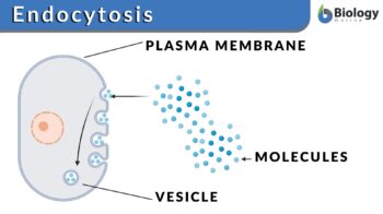
In the process of endocytosis, the cell membrane folds itself around the material to be engulfed resulting in the formation of a vesicular structure that eventually pinches off from the membrane inside the cell.
It is important to understand whether endocytosis is an active or passive process. Does the endocytosis process require energy? Endocytosis is an active process. That means ATP molecules are involved in this process. Macrophages engulf the pathogenic material (e.g. viruses or bacteria) and eventually neutralize them with the help of lysosomes. The endocytosed material is digested with the help of hydrolytic enzymes of the lysosomes.
Endocytosis is employed by the cells in order to:
- Procure the nutrients for cellular growth and repair,
- Seize the toxin or unwanted pathogens and eventually neutralize them in the cells
- Eliminate the old or non-functional cells
Endocytosis is a process in which a cell takes in materials from the outside by engulfing and fusing them with its plasma membrane. Etymology: Greek éndon, meaning “within”, + “kutos”, meaning “hollow vessel” + -“osis”, expressing state or condition. Compare: exocytosis.
An opposite cellular phenomenon wherein the cell expels out the material from its interior to the external environment is known as exocytosis.
So, what are exocytosis and endocytosis? Endocytosis and exocytosis are the processes responsible for the bulk transportation of the cellular material. Exocytosis is the process by which cells eliminate or release the material into extracellular space. In the process of exocytosis, during the fusion of a vesicle with the plasma membrane cellular content is transferred into the extracellular space. The exocytosis process is required for the removal of the cellular undesired /waste material, for cellular communication, for enabling cellular growth and repair. This is the usual mechanism employed by macrophages to eliminate waste or pathogenic material. Examples of exocytosis include glucagon transportation from the pancreas to the liver, the release of neurotransmitters into the synaptic cleft, etc.
The major difference between the two modes of cellular transport — endocytosis and exocytosis — are enlisted in Table 1.
Table 1: Endocytosis vs. Exocytosis |
|
|---|---|
| Endocytosis | Exocytosis |
| Endocytosis (biology definition) is the process of engulfing the material from the external environment into the cell | Exocytosis (biology definition) is the process of eliminating the material from the cell into the external environment |
| Cell membrane folds and forms a vesicular structure around the material to be ingested | The secretory vesicle fuses with the plasma membrane |
| The primary purpose is to:
Absorption of nutrients, eliminating old cells and neutralizing pathogenic material |
The primary purpose is to:
Eliminating cellular waste, mode of cellular communication, and cellular repair |
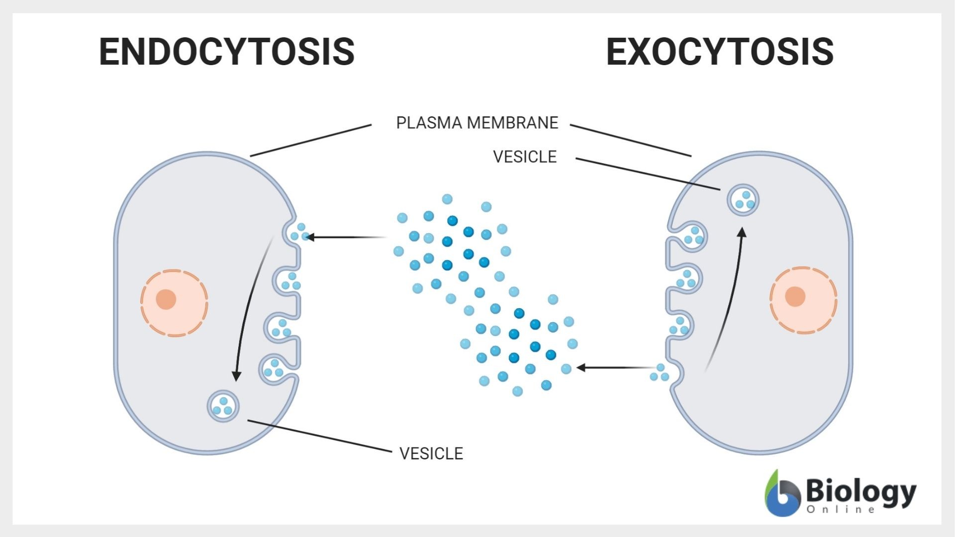
Endocytosis pathway overview
- Invagination: Cellular membrane folds and creates a cavity wherein the material to be engulfed is entrapped.
- Vesicle formation: The cellular membrane folds back forming a vesicle with uniform vesicular membrane thickness. Material to be engulfed is present inside this vesicle.
- Once the vesicle is formed, it detaches from the cellular membrane and proceeds for further processing by the cell.
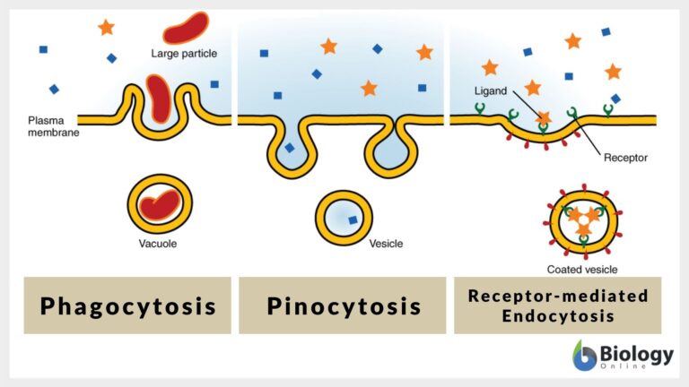
Types of Endocytosis
What are the types of endocytosis? The different types are as follows: (1) phagocytosis, (2) pinocytosis, (3) receptor-mediated endocytosis, and (4) caveolae.
Phagocytosis
Phagocytosis or cell eating is a type of endocytosis wherein large cells ingest large foreign particles or material. The cell which undertakes this endocytosis is known as a phagocyte. The phagocytosis process was discovered in 1882 by Élie Metchnikoff.
What is phagocytosis?
Phagocytosis (definition in biology) is the cellular uptake process by which a cell ingests or engulfs the material of large size with the modulation of the plasma membrane.
Phagocytosis was first discovered in 1867 by a Canadian physician, William Osler. Macrophages, monocytes, neutrophils, eosinophils, and dendritic cells are some of the white blood cells that exhibit phagocytosis.
Phagocytosis is a five-step process:
- Detection: It is the first step of phagocytosis wherein; the phagocyte recognizes the foreign substance or the antigen and starts the movement towards the target substance.
- Attachment: The phagocytic cell attaches itself to the target. As a result of this binding pseudopodia formation is initiated. Pseudopodia is the extended cell membrane that surrounds the target.
- Ingestion: The membrane of pseudopodia fuses to form a vesicle and the target material is in the vesicle. This vesicle wherein the target is enclosed is known as a phagosome.
- Fusion: The phagosome fuses with the lysosome to form a phagolysosome. Lysosomes contain different hydrolytic enzymes. These hydrolytic enzymes digest the materials present in the vesicle.
- The digested material is eliminated from the cell by the process of exocytosis.
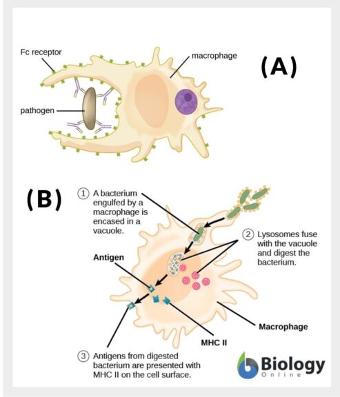
Examples of phagocytosis
Immune cells such as macrophages, dendritic cells, and neutrophils exhibit phagocytosis for neutralizing and eliminating the pathogenic material from the body. A white blood cell engulfing a bacterium is a classic example of phagocytosis. The largest immune cells are the macrophages that specialize in detecting the antigen, followed by attaching to it leading to ingestion and fusion resulting in neutralizing the antigenic material and eventually eliminating it by the process of exocytosis. Macrophages have pseudopodia and are the major immunogenic cells in the body. The antigen is the substance, which the body recognizes as foreign and undesirable to the body. It can be viruses, bacteria, fungi, pollen grains, etc. For the process of adherence, sometimes a phagocytic cell needs certain specific protein components from the blood that forms a layer over the antigen to be engulfed. These proteins are known as Opsonin and this process of forming a layer over the antigen is known as opsonization. Once opsonization is complete, the phagocytic cell then engulfs the antigen by the process of phagocytosis.
Watch the video below showing phagocytosis under a microscope (a macrophage phagocytosing a bacterial cell).
Pinocytosis
Pinocytosis or cell drinking (or endocytosis of fluid) is the process wherein the cell engulfs or ingests the small particles of the fluid.
What is pinocytosis?
Pinocytosis definition in biology is the process of cellular uptake or ingestion of fluid or particles of small size by a cell with the modulation of the plasma membrane.
The process of pinocytosis differs from phagocytosis in the formation of pseudopodia. In pinocytosis, the formation of pseudopodia is absent. Rather, there is the only formation of vesicles. The process of pinocytosis follows basic steps of the process of endocytosis wherein a vesicle is formed around the small particles or fluid to be engulfed. The vesicles are then internalized in the cell and may fuse with the cellular lysosome. Eventually, the contents are digested by the hydrolytic enzymes of the lysosomes.
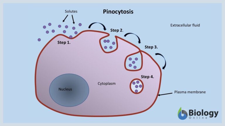
Types of pinocytosis
Pinocytosis can be of two types:
- Micropinocytosis: vesicles of small size (0.1µm in diameter) are formed. The vesicles bud off from the cell and most of the body cells exhibit micropinocytosis.
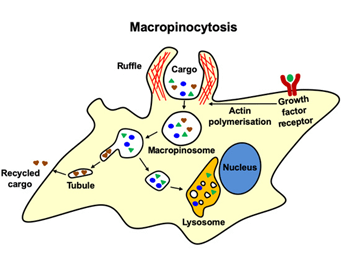
- Macropinocytosis: vesicles of large size (0.5 to 5 µm in diameter) are formed. However, vesicles are not formed by the process of budding off from the cell. In micropinocytosis, plasma membrane ruffles or extends into the extracellular space to engulf the material to be ingested and eventually roll back into the intracellular space to form vesicles. This process is widely exhibited by white blood cells.
Pinocytosis examples
The following are examples of pinocytosis:
- Uptake of hormones and enzymes by the body cells
- Uptake of nutrients from the surrounding environment by human egg cells
- Uptake of nutrients by the microvilli cells of the intestines
Receptor-mediated endocytosis
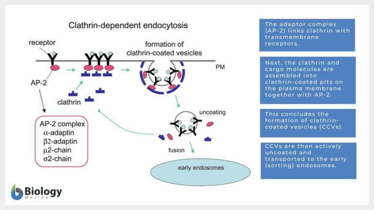
This endocytosis is also known as clathrin-mediated endocytosis. As suggested by the name, this endocytosis process is facilitated through a coat protein known as clathrin. This is one of the best-studied endocytic processes. The molecules, that are to be engulfed, fuse with specific receptors on the cell membrane, which are found in the protein clathrin-rich regions of the plasma membrane. These regions are known as clathrin-coated pits. After receptor binding of the molecule is carried out, clathrin pits undergo the process of internalization forming clathrin-coated vesicles. These clathrin-coated pits then fuse with the endosome which results in the removal of the clathrin coat from the internalized vesicles. Here we need to understand what an endosome is. Endosomes are the membrane-bound intracellular cell organelle in the eukaryotic cells. The function of endosomes is to carry out the sorting of the material that has been internalized and its delivery to the lysosomes.
Once, clathrin is removed, these vesicles are then digested upon. This process ensures the uptake of specific material like metabolites, hormones, proteins, and certain types of viruses. Thus, the key difference between pinocytosis and receptor-mediated endocytosis is that the pinocytosis endocytosis of the non-specific molecules is carried out by the cell whereas in receptor-mediated endocytosis uptake of specific molecules occurs.
Essential steps of Receptor-mediated Endocytosis or Clathrin-Mediated Endocytosis
- The molecule to be engulfed binds to specific receptors on the plasma membrane and this complex then drifts to the region that contains a clathrin-coated pit.
- These molecule-receptor complexes collect in the clathrin-coated pit, which eventually invaginates and forms a vesicle.
- Eventually, a clathrin-coated vesicle containing the molecule-receptor fuses with the cellular endosome leading to the removal of the clathrin coating and the receptor molecule. Most of the time, the receptor molecules are recycled back to the plasma membrane.
- Once the clathrin coating is removed, the vesicle, then, fuses with lysosomes wherein the enzymes degrade the vesicular content and deliver the required contents back to the cytoplasm.
Receptor-mediated endocytosis is a more efficient and selective endocytic process than pinocytosis. The classical examples of receptor-mediated endocytosis are uptake of cholesterol-bound low-density lipoproteins, recycling of iron-bound transferrin, and the chief endocytic process in plant cells. Hypercholesterolemia is the result of the defective or absent low-density lipoprotein receptors. As a result of this, cholesterol is not eliminated from the body and is deposited in the blood vessels resulting in atherosclerosis.
Caveolae
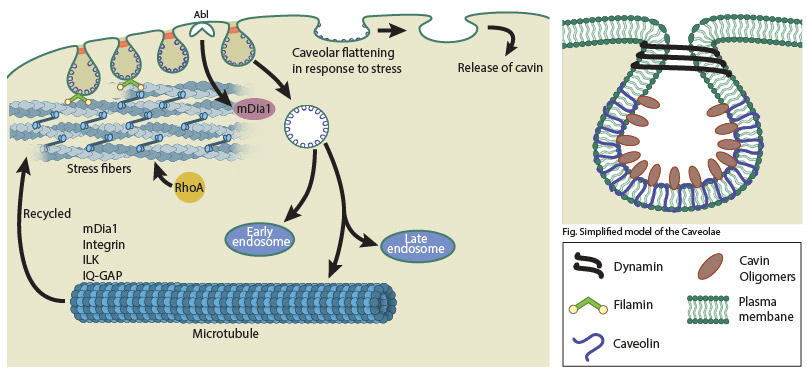
This is a clathrin-independent, receptor-mediated- cholesterol, and dynarrin- dependent pathway endocytic process wherein invagination of the plasma membrane results in the formation of bulb-shaped cavities inside the cells which are known as caveolae. The literal meaning of the word ‘caveolae’ is ‘little caves’. The size of these cavities is 50-80nm.
Two proteins, i.e., caveolins and cavins, facilitate the process of formation of caveolae. Caveolins are an integral protein of 21 kDa of the membrane whereas cavins are the peripheral proteins of the membrane. Three caveolins, i.e., caveolin-1, caveolin-2, and caveolin-3 are involved in the formation and stability of the caveolae. Caveolin-3 is a specific protein found only in muscle cells. These three caveolin work in conjunction with four types of cavins (cavin-1, -2, -3, and -4; cavin-4 is specifically found in muscle cells) to form caveolae.
Caveolae are completely absent in neurons while these are most widely found on the endothelial cells. Palade and Yamada first reported caveolae in the 1950s.
Function of Endocytosis
Endocytosis is basically a bulk transportation mechanism of the cells.
- Receptor signaling
- Uptake of nutrients
- Remodeling of the plasma membrane
- Controlling and neutralizing the entry of pathogens
- Neurotransmission of the essential signals
- Modulating response of the cells towards various cell signals
Endocytosis Examples
Transportation and endocytosis of the hydrophobic molecules like, steroids, retinol, etc. by binding with soluble proteins. It has been found that vitamin A (retinol) is bound to the retinol-binding protein, vitamin D3 is bound to the vitamin D-binding protein, cortisol is bound to the corticosteroid-binding globulin, and testosterone and estrogens bound to the sex hormone-binding globulin are endocytosed via receptor-mediated endocytosis.
Similarly, the removal of hydrophobic and insoluble cholesterol from the body is facilitated by its soluble complex with low-density lipoproteins through the process of receptor-mediated endocytosis. Thus, defective or absence of low-density lipoprotein receptors results in the accumulation of cholesterol leading to hypercholesterolemia and atherosclerosis, both of which can eventually lead to a heart attack.
Although, the body utilizes the process of endocytosis to neutralize the pathogens. There are certain pathogenic intracellular parasites that utilize endocytosis, specifically receptor-mediated endocytosis, to gain access and establish themselves intracellularly in the host cell. Epstein-Barr Virus (EBV), Influenza virus, Listeria monocytogenes, and Streptococcus pneumonia are some of the pathogens that utilize receptor-mediated endocytosis by tricking the immune system and establish themselves in the host cells.
Try to answer the quiz below to check what you have learned so far about endocytosis.
References:
- Miaczynska, M., & Stenmark, H. (2008). Mechanisms and functions of endocytosis. Journal of Cell Biology, 180 (1): 7–11. doi: https://doi.org/10.1083/jcb.200711073.
- Lin, X.P., Mintern, J.D., Gleeson, P.A. (2020). Macropinocytosis in Different Cell Types: Similarities and Differences. Membranes 10, 177.
- Wu, F., & Yao, P. J. (2009). Clathrin-mediated endocytosis and Alzheimer’s disease: an update. Ageing research reviews, 8(3), 147–149. https://doi.org/10.1016/j.arr.2009.03.002
- Kiss A. L. (2012). Caveolae and the regulation of endocytosis. Advances in experimental medicine and biology, 729, 14–28. https://doi.org/10.1007/978-1-4614-1222-9_2’
- Pelkmans, L. and Helenius, A. (2002), Endocytosis Via Caveolae. Traffic, 3: 311-320. https://doi.org/10.1034/j.1600-0854.2002.30501.x
©BiologyOnline.com. Content provided and moderated by BiologyOnline Editors.

