Endomembrane System
n., plural: endomembrane Systems
[ˈɛndəʊˈmembɹeɪn ˈsɪstəm]
Definition: a system of membranous components involved in biomolecular synthesis and transport
Table of Contents
Ever wondered how biomolecules are made within the cell and then they are released outside the cell for use by the body? Let’s take, for example, lipids and hydrolytic enzymes that are released by the lamellar bodies. These biomolecules are released into the skin so that the skin would shed its “dead” outermost layer. How about other proteins, like keratins? Keratins are the fibrous proteins present in hair, nails, skin, and many other parts of our body.
How does our body produce these biomolecules? Proteins, carbohydrates, lipids, and nucleic acids are the biomolecules that are crucial to our daily functions. They play an essential role in growth, reproduction, digestion, immune defense, and homeostasis. Without them, life will cease to exist.
Thus, these biomolecules have to be made, as well as regulated, to enable and sustain life. Depending on what’s needed, your cells produce multifarious biomolecules through various organelles working as a single unit. For this, they are referred to altogether as the “endomembrane system”.
What Is the Endomembrane System?
To define the endomembrane system, we should first be familiar with the term, “organelle” or “little organs”. An organelle refers to the various structures of the cell that perform a particular function. An example of an organelle is a nucleus, which is the organelle of the cell that directs cell activity, and therefore acts as the cell’s control center.
A stricter definition of an organelle, though, is that the cell structure must be a compartment or a “sac”, which means a biological membrane surrounds the contents to separate them from the outside. With this definition, an example of structures inside the cell that are not membrane-bound is the ribosomes.
Nonetheless, other references consider ribosomes as organelles, specifically as a non-membrane-bound type (as opposed to the membrane-bound). Nevertheless, the ribosomes are not part of the endomembrane system. And so you might ask, which organelles, therefore, comprise the endomembrane system of a cell? And which structure is not part of the endomembrane system? To answer that, let’s get to know the different endomembrane system parts.
What are the different parts or components of the endomembrane system? Look at Figure 1. That is a typical cell of a eukaryote. A eukaryotic cell is a type of cell that has a nucleus and other membrane-bound organelles. The presence of membrane-bound organelles is used to identify a eukaryotic cell from a prokaryotic cell.
A prokaryotic cell lacks membrane-bound organelles. You won’t find a nucleus inside a prokaryotic cell, such as a bacterial cell. Prokaryotes lack an intra-membrane system. Conversely, a eukaryotic cell has an internal membrane system that separates and compartmentalizes cell contents.
Therefore, the endomembrane system characterizes a eukaryotic cell; it is absent in a prokaryotic cell. A human cell is an example of a eukaryotic cell.
Although the biomolecular contents of the organelles are separated by biological membranes, certain biomolecules may be transported from one organelle to another. The eukaryotic cells are able to move their organelles’ biomolecular contents via the endomembrane system that connects the component organelles.
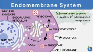
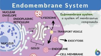
Endomembrane system is a system of membranes within a cell that serves as a single functional and developmental unit. The endomembrane system is a system of membranous components. It includes the membranes of the nucleus, the endoplasmic reticulum, the Golgi apparatus, lysosomes, endosomes, vesicles, and the cell membrane. It does not include the membranes of mitochondria and/or chloroplasts.
Endomembrane System Function
What is the function of the endomembrane system? In general, it is involved in the creating and distributing of the newly-made biomolecules. The nuclear envelope has holes through which the mRNA transcript (code for creating protein) passes through. The endoplasmic reticulum where ribosomes are attached is associated with the production of proteins whereas the part of the ER where ribosomes are not attached to its surface serves as the site for lipid and carbohydrate syntheses as well as for calcium ion storage.
The Golgi apparatus is the packaging site of the cell. It “packs” the newly synthesized biomolecules for transport within or outside the cell. The lysosomes contain digestive enzymes for intracellular digestion. The digestive enzymes are produced from the ER and released from the Golgi apparatus.
The endosomes are involved in the endocytic membrane transport pathway by which the molecules from the cell membrane are taken into the lysosome.
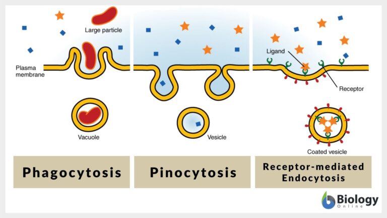
The cell membrane is the protective barrier that separates the interior of the cell from the outside environment. It is also involved in cell-cell contact and signaling. It is also responsible for taking in material from the outside into the cell (endocytosis) and for moving materials from the cell to the outside (exocytosis).
Know the difference between: Endocytosis and Exocytosis
Components of the Endomembrane System
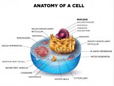
Take a look at the schematic diagram of an animal cell below (Figure 1). The different parts are as follows: (1) nucleolus, (2) nucleus, (3) ribosomes, (4) vesicle, (5) rough endoplasmic reticulum, (6) Golgi apparatus, (7) cytoskeleton, (8) smooth endoplasmic reticulum, (9) mitochondrion, (10) vacuole, (11) cytosol, i.e. the fluid that contains organelles, comprising the cytoplasm, (12) lysosome, (13) centrosome, (14) cell membrane. Of these 14 parts labeled in this diagram, only seven are components of the endomembrane system:
- Nuclear envelope (#2)
- Endoplasmic reticulum (#s 5 & 8)
- Golgi apparatus (#6)
- Lysosomes (#12)
- Endosomes (not shown in the diagram)
- Vesicles (#4)
- Plasma membrane (or Cell membrane) (#14)
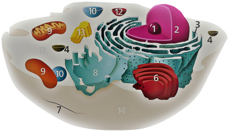
Their membranes are related through direct or indirect contact. By direct contact, it is exemplified by the nuclear envelope being connected to the membrane of the endoplasmic reticulum (ER). The membrane of the ER, in turn, is connected to the Golgi apparatus. By indirect contact, an example would be is the vesicle forming by taking membrane segments from the plasma membrane.
Other membrane-bound organelles, such as mitochondria and chloroplasts (Note: chloroplasts are not present in an animal cell but in a plant cell), are not included in the endomembrane system because they are not in any way in contact with them. Both mitochondria and chloroplasts, are in fact, regarded as “semi-autonomous organelles” as they have their own DNA.
Let’s, now, take a look further at the different components of the endomembrane system.
1. Nuclear Envelope
One of the most prominent organelles in a eukaryotic cell is the nucleus. It is the large, membrane-bound structure that contains the genetic material (DNA) that may organize into chromosomes. Its biological membrane has a special name — the nuclear envelope (also called nuclear membrane). The nuclear envelope is made up of two lipid layers. It has holes called “nuclear pores”.
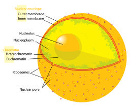
So this is where protein synthesis begins. The DNA segment that carries the code for a particular protein is copied via the process called transcription. In essence, it is called transcription because it produces a “transcript” in the form of mRNA. This transcript is actually a copy of the “code” when making a protein. It was copied from the specific coding region of the DNA.
The newly synthesized transcript (mRNA) leaves the nucleus through the nuclear pore. The export receptors in the nuclear membrane guide the mRNA out of the nucleus through a nuclear export signal added to the mRNA during transcription. Once in the cytosol, the nuclear export signal is taken off from the mRNA and then it returns to the nucleus. (Ref. 1) (See Figure 3)
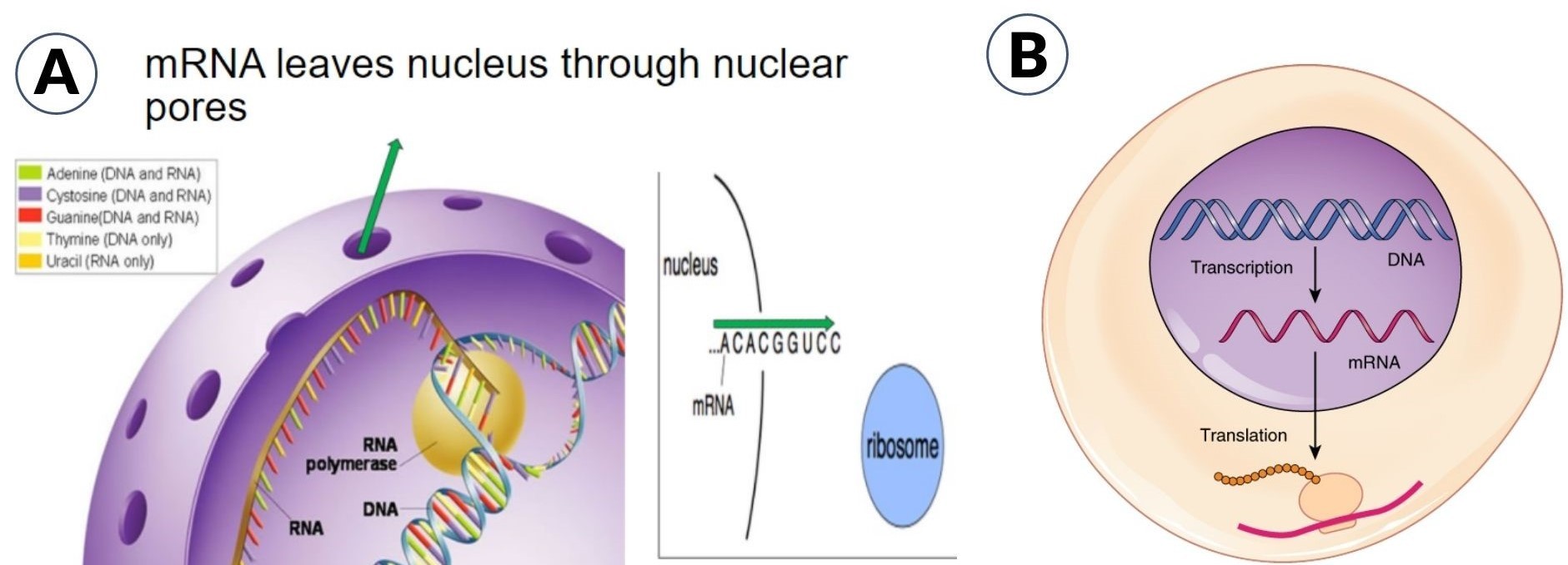
In the cytosol, the mRNA is identified by the ribosome for translation. It translates the code into amino acids with the help of matching tRNAs.
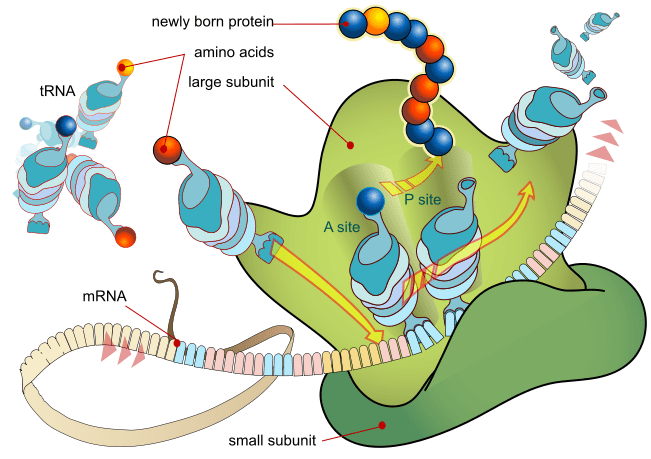
One of the possible scenarios after translation is that it will be taken into the endoplasmic reticulum for maturation. The newly-formed protein has to go through protein folding or post-translation modifications to become “mature” proteins. The cytosol is reductive as opposed to the oxidative lumen (biology definition: the fluid-filled cavity within the endoplasmic reticulum and the Golgi apparatus). This means that there are post-translational steps, such as disulfide bond formation, that would rather occur inside the lumen of these organelles (which is oxidative) rather than in the cytosol (which is reductive). (Ref. 2)
2. Endoplasmic reticulum (ER)
What is an endoplasmic reticulum? What does it do? If the nucleus is the first site of protein synthesis, the endoplasmic reticulum (ER) acts as the first site of the secretory pathway. In Figure 5, the location and structure of the endoplasmic reticulum are shown. Take note that the outer membrane of the nuclear envelope is continuous with the ER.
There are two types of ER: (1) rough endoplasmic reticulum (rough ER) and (2) smooth endoplasmic reticulum (smooth ER). Rough ER has ribosomes attached to its surface whereas smooth ER has no ribosomes.
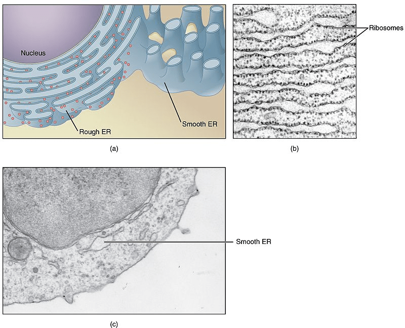
Rough ER
What does the rough ER do? As mentioned above, the ribosomes are where proteins are made. They pick up mRNAs to translate their code into a new protein. While there are ribosomes in the cytosol, there are also ribosomes that are attached to the ER — the latter defines the ER as a “rough”-type. Because of the presence of ribosomes, the rough ER, therefore, is associated with the production of proteins.
After translation, some of the new proteins are moved into the ER’s lumen for protein folding or post-translational modifications. This is called “post-translational translocation”. In another event called “cotranslational translocation”, the ribosome takes the initial chain of amino acids (peptide) to the ER even without finishing the translation yet.
Cotranslational translocation occurs when the ribosome hits about 16-30 amino acids that are recognized altogether by the signal recognition particle as a signal peptide. The signal peptide is often composed of a series of hydrophobic acids after one positively charged amino acid. (Ref. 2)
The ribosome, together with its peptide cargo, moves to the ER and docks to the surface by binding to the ER surface (via the binding site called translocon). The binding calls for GTP molecules, which then would attach to them, strengthening the interaction. This activates ER membrane protein complex to form a passageway (translocation channel) through which the peptide can pass through and reach the ER lumen. The ribosome, then, resumes translating the mRNA.
As more and more amino acids are added to the signal peptide, the peptide is pushed into the ER lumen through the translocation channel. After translation, the whole protein is eventually released into the ER lumen. The signal peptide is cleaved off by a signal peptidase. (Ref. 2) The nascent protein will then undergo maturation.
That’s for proteins but what about other biomolecules like lipids. Where are lipids made in the cell? Let’s find out below.
Smooth ER
The smooth ER is part of the endoplasmic reticulum that lacks ribosomes. As described above, the rough ER is that part wherein ribosomes are bound. After translation, though, the ribosomes leave the surface and that becomes a “smooth” ER again. If no ribosomes are attached to it, what does it become then? It becomes the site of other biosyntheses, particularly lipid synthesis. This is where lipids are made, such as phospholipids, sterols, steroids, ceramides, and triglycerides. For example, in triglyceride synthesis, three fatty acids are esterified to glycerol in the smooth ER lumen. The presence of various enzymes enables these biosyntheses.
Apart from lipid synthesis, what else does smooth ER do? The smooth ER that regulates intracellular calcium concentration has a special name. It is called a sarcoplasmic reticulum and it is found in muscle cells. Therefore, this type of smooth ER is associated with muscle movement.
It is also associated with carbohydrate metabolism. Glucose, the primary source of energy, can be derived from other sources apart from dietary carbohydrates. Smooth ER contains the enzyme, glucose-6-phosphatase, which converts glucose 6-phosphate into glucose.
Smooth ER is also where drug detoxification occurs. Liver cells, in particular, have cytochrome P450s residing in the smooth ER lumen. These enzymes help detoxify drugs and poisons, for example, by adding a hydroxyl group to the drug molecule.
For the summary of the two types of ER, their definition, structure, and function, refer to Table 1.
Table 1: Rough ER vs. Smooth ER | |
|---|---|
| Rough Endoplasmic Reticulum | Smooth Endoplasmic Reticulum |
| Rough ER definition: an organelle made up of interconnected flattened sacs with ribosomes attached to the surface | Smooth ER definition: an organelle made up of interconnected flattened sacs, with no ribosomes bound on its surface |
| Rough ER structure: Consists of tubules (tubular) and vesicles (rounded sacs) that are arranged in a reticular pattern. The outermost is a membrane and the innermost is a cavity called lumen that is fluid-filled. | Smooth ER structure: Similar to rough ER but devoid of ribosomes docked on the outer surface of the membrane |
| Rough ER function: involved in protein synthesis and site of cotranslational translocation | Smooth ER function: lipid synthesis, calcium ion storage and regulation, carbohydrate metabolism, drug detoxification |
Watch this vid to see an endoplasmic reticulum cartoon and other animated images of what’s inside the cell. Drag the screen while watching!
3. Golgi Apparatus
The Golgi apparatus (also called Golgi complex or simply Golgi) is an organelle that, similar to ER, is made up of cisternae (flattened membrane sacs containing fluid). In animal cells, Golgi cisternae are connected by microtubules; in plant cells, they are connected by actin. And unlike the endoplasmic reticulum, the Golgi cisternae are not connected directly to the nuclear envelope. Nevertheless, the Golgi cisternae came from the vesicles that bud off from the endoplasmic reticulum. Thus, a part of the Golgi apparatus is commonly seen near the endoplasmic reticulum exit sites. (Ref. 3)
The Golgi apparatus is made up of cisternae forming a stack. Depending on the location of the cisternae in the stack, they may be cis, medial, or trans. Each of them possesses specific enzymes anchored in their membrane, and therefore involved in specific biological activities. In essence, the cis face contains enzymes that are involved in the early modifications of proteins whereas the trans face contains enzymes for final protein modifications.
These cisternae are not fixed to their positions. They move outward. Thus, the cis face cisternae are found closest to the ER. The medial is the central cisternae. The trans face cisternae are farthest from the ER. That means a cisterna starts out as cis, then, becomes medial, and ultimately, trans, with each stage possessing new and different sets of enzymes as it moves away from its starting point. It carries and modifies the protein that it had from the start. Instead of emptying its protein content into another cisterna, it keeps the protein, modifying it until it reaches its final “mature” state. (Ref. 2) Therefore, the correct order of movement of proteins through the Golgi apparatus is from cis– to trans–.
So, what does the Golgi apparatus do? What is the function of the Golgi apparatus? In terms of protein synthesis, the Golgi apparatus function is to modify protein until it becomes “mature” and ready for secretion. Its primary function is to serve as the “packaging center of the cell”. For example, it sorts the proteins coming from the ER, and then tags the proteins to their destination sites.

The biomolecules inside the Golgi vesicle typically will have one of these fates:
(1) for exocytosis
(2) for storage and later secretion(e.g. as secretory vesicles)
(3) for intracellular transport
(4) for degradation (either as a new lysosome or for fusion with the existing lysosome)
4. Vesicles
What is a vesicle? In general, a vesicle is a small sac. But what about in cell biology — what are vesicles? Inside the cell, the vesicles refer to any bubble-like structures that store and transport cell products within the cell. Its contents are separated from the cytosol by at least one lipid bilayer.
There are different vesicles inside the cell. The ER vesicles, for instance, are the transport vesicles that pinch off from the ER to translocate the protein cargo, for example, to the cis face of the Golgi apparatus. Another transport vesicle is the Golgi vesicle, which in turn, is defined as the vesicle that buds off from the Golgi to transport its cargo either internally (via intracellular transport) or externally (via exocytosis or by secretion as secretory vesicles).
Lysosomes are vesicles that digest metabolic wastes. Another example of vesicles is vacuoles. The function of the vacuoles is generally for osmoregulation. For more info about transport vesicle function, see Figure 7.

5. Lysosomes
What is a lysosome? A lysosome refers to the membrane-bound cell structure that contains digestive enzymes. And so, what does a lysosome do? The digestive enzymes inside the lysosome are used in “digesting” worn-out organelles, misfolded proteins, engulfed viruses or prokaryotes, and food particles. The lysosomes are also involved in cell membrane repairs. If the cell is beyond repair, the lysosomes will “self-destruct” and so the reason why they are referred to as “suicidal bags”. Their contents are very acidic and the digestive enzymes (hydrolytic enzymes) will break down large complex molecules into smaller, simpler molecules. See Figure 8 for lysosome structure and function.
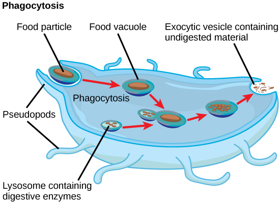
Lysosomes vs. Peroxisomes: Peroxisomes are cytoplasmic structures that breakdown a very long chain of fatty acids, polyamines, and D-amino acids by beta-oxidation. While they can be easily mistaken as lysosomes, peroxisomes are cytoplasmic structures with a different function, and most importantly, they are not part of the endomembrane system.
6. Endosomes
Endosomes are membrane-bound cytoplasmic structures through which molecules that have been taken into the cell via endocytosis (see Figure 9) pass en route to the lysosome for “digestion” (see Figure 10). Similar to lysosomes, the endosomes are single-membraned.

7. Cell Membrane
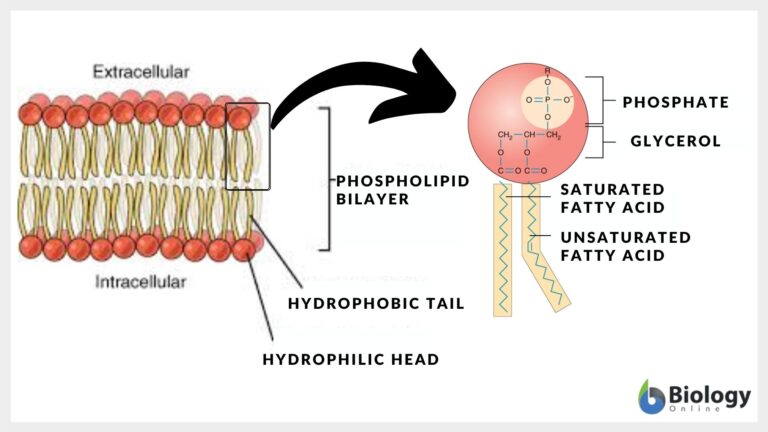
The cell membrane is a membrane surrounding the protoplasm (the living component of the cell). Both eukaryotic and prokaryotic cells have a cell membrane that separates the protoplasm from the outside environment. Now, what is the main component of the cell membrane? The cell membrane is made up of two layers of lipids, chiefly, phospholipids.
In Figure 11, the phospholipids of the cell membrane are arranged in a way that their hydrophilic heads are facing outward while their hydrophobic tails are pointing inward. This organization is essential in making the cell membrane “selectively permeable”. It means that the cell membrane is permeable to select particles as not all of them will be allowed to pass through.
See Figure 12. Also present in the cell membrane are proteins, glycoproteins, sterols, and glycolipids (lipids with carbohydrates).

READ: Movement of Molecules Across Cell Membranes
Tracking Newly-made Biomolecule: Keratin and Ceramides (Example of Endomembrane System Biosynthetic Pathway)
Now that we learn about the various endomembrane system components and their functions let’s track the order of movement of proteins through the endomembrane system after translation.
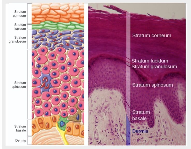
Going back to keratin, hydrolytic enzymes, and lipids as our example, these biomolecules are produced by specialized cells called keratinocytes that are located on the outermost layer of our skin (epidermis). In essence, these cells produce copious amounts of keratins inside them, arranged in parallel bundles. See Figure 13. This helps create a protective barrier against heat, water loss, irritants, allergens, microbial assaults, and other stressors from the environment.
During cornification (protective barrier formation), the keratinocytes on the topmost layer of the epidermis produce more and more keratin. This process is called keratinization. Eventually, the keratinocytes lose their nucleus and other organelles. As a result, metabolism ceases, and eventually, they become almost filled with keratin. At this point, the “dead” (terminally differentiated) keratinocytes are referred to as corneocytes (also called squames).
These corneocytes are interlocked with one another to form a physical barrier referred to as the stratum corneum (the topmost layer of the epidermis). The corneocytes have also replaced their cell membrane with a cornified cell envelope. (Ref. 5,6)

Now, since these cells have reached their ultimate fate and are no longer “living”, they are periodically shed and replaced by newer keratinocytes from the deeper layers of the epidermis. They went up to replace squames. So, let’s track some of the biomolecules involved. Let’s begin!
When the keratinocytes move up to the topmost layer of the skin, they will differentiate into corneocytes by undergoing keratinization. The cells will be filled with keratin. Prior to the disintegration of the nucleus, the cell will synthesize keratin proteins based on the genetic code in the DNA in the nucleus.
In human skin, the keratin is a complex of type I and type II alpha-keratins, which are encoded on chromosomes 17 and 12, respectively. (Ref. 7) Inside the nucleus, the copy of the codes for type I and type II from these genetic locations are made through transcription. mRNA (transcripts) are made. These mRNAs carrying the codes leave the nucleus to travel to the ribosomes in the cytosol. tRNAs “translate” the code from the mRNA by bringing in the correct amino acids that match the code. Type I and type II form a keratin complex (called “coiled-coil”). As more and more coiled-coil dimers are formed, they bond together via disulfide bonds, and align to form a protofilament. An aggregate of two protofilaments forms a protofibril and then four protofibrils form an intermediate filament, which, in this regard is alpha-keratin. These keratin filaments will, then, connect the cell to the adjacent cell via desmosomes. In Figure 13:B, desmosome components, desmoplakin and plakoglobin, anchor the keratin filaments between cells via desmosomal plaques arranged on the lateral sides of the plasma membranes. Take note that the keratin just described is not for cellular secretion. Because of that, these alpha-keratins stay inside the cell and do not enter the secretory pathway.
During the early translation, the code for alpha-keratins does not include a signal peptide and so it is likely translated in the ribosomes in the cytosol. Nevertheless, it is likely that the dimers form disulfide bonds in the ER as the disulfide bond formation post-translation typically occurs in the lumen of the ER as explained in the earlier section. And then for further maturation, the Golgi apparatus is the likely site.
As for the lipids, proteins, and hydrolytic enzymes inside the lamellar bodies, these biomolecules are for secretion. If you will recall, the lamellar bodies (also called keratinosomes or Odland bodies) are special vesicles (secretory organelles) that contain biomolecules that have to be taken outside the cell to help the skin shed its “dead” outermost layer. This natural and periodical peeling of our skin is called desquamation.
Keratinocytes in the stratum spinosum and stratum granulosum (see Figure 14) have lamellar bodies that contain various cargoes, such as lipids (e.g. glucosylceramides), hydrolytic enzymes (e.g. proteases, lipases, etc.), and several other proteins (e.g. corneodesmosin). (Ref. 8) These proteins are encoded by specific genes in the nuclear DNA. Corneodesmosin, for instance, is encoded by the CDSN gene in chromosome 6 of humans. (Ref. 9)
After copying the DNA codes into mRNA transcripts, the transcripts are translocated from the nucleus into the cytosol where ribosomes pick them up for translation. Since these proteins are for secretion, they enter the secretory pathway.
mRNA for corneodesmosin, in particular, encodes a 539-amino acid protein with an N-terminal signal peptide and one putative N-glycosylation site (which indicates that it is indeed for secretion and it is glycosylated. (Ref. 10) It is, therefore, shuttled by the ribosome to the rough ER for further translation. Then, it is shipped to the cis face of Golgi for further maturation until such time that it reaches the trans face (exit point) for secretion.
As for lipids, they are synthesized in the smooth endoplasmic reticulum. The products are then transported into the Golgi apparatus in a similarly cis-to-trans direction. When mature, the cargoes are packaged by the Golgi apparatus and dispatched to their destination, and in this example, to the lamellar body.
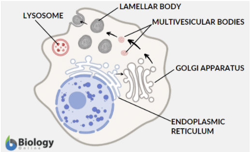
Electron microscopy studies revealed that the lamellar bodies are branched, tubular vesicles derived from the trans-Golgi. Also, research findings indicate that the components of the lamellar bodies seem to be delivered via independent shuttling of various cargoes through multivesicular bodies. And because of the presence of hydrolytic enzymes and other features similar to the lysosomes, the lamellar bodies are suggested to be a special kind of lysosome. (Ref. 11) (Figure 15)
Takeaways: What is theEndomembrane system and its function? The membranes of the organelles included in the endomembrane system are related through (1) direct contact: for example, the nuclear envelope is connected to the endoplasmic reticulum, and the endoplasmic reticulum, to the Golgi apparatus and (2) indirect contact: for example, by the transfer of membrane segments as vesicles. The endomembrane system is involved in the manufacturing and distribution of cellular products. Nonetheless, the membranes of the organelle components vary in specific functions. For instance, the nuclear envelope encases the nuclear material. The endoplasmic reticulum is associated with the synthesis of proteins and other biomolecules. The Golgi apparatus does the packaging of newly synthesized biomolecules for transport within or outside the cell. The lysosomes are vesicles containing enzymes synthesized from the endoplasmic reticulum and released from the Golgi apparatus. Endosomes are compartments of the endocytic membrane transport pathway from the cell membrane to the lysosome. The cell membrane is the protective barrier that separates the interior of all cells from the outside environment.
Try to answer the quiz below to check what you have learned so far about endomembrane system.
References
- Alberts, B., Johnson, A., Lewis, J., Raff, M., Roberts, K., & Walter, P. (2021). The Transport of Molecules between the Nucleus and the Cytosol. Nih.gov; Garland Science. https://www.ncbi.nlm.nih.gov/books/NBK26932/
- Minikel, E. V. (2013). Cell Biology 04: The Secretory Pathway. Cureffi. https://www.cureffi.org/2013/02/24/cell-biology-04-the-secretory-pathway/
- Suda Y, Nakano A (April 2012). “The yeast Golgi apparatus”. Traffic. 13 (4): 505–10.
- Do Plant Cells have Lysosomes? (2021). Millerandlevine.com. http://www.millerandlevine.com/ques/lysosomes.html
- Haftek, M., Callejon, S., Sandjeu, Y., Padois, K., Falson, F., Pirot, F., Portes, P., Demarne, F., & Jannin, V. (2011). Compartmentalization of the human stratum corneum by persistent tight junction-like structures. Experimental Dermatology, 20(8), 617–621. https://doi.org/10.1111/j.1600-0625.2011.01315.x
- Scott, D. W., & Miller, W. H. (2011). Structure and Function of the Skin. Equine Dermatology, 1–34. https://doi.org/10.1016/b978-1-4377-0920-9.00001-9
- Lee, C.-H., & Coulombe, P. A. (2009). Self-organization of keratin intermediate filaments into cross-linked networks. Journal of Cell Biology, 186(3), 409–421. https://doi.org/10.1083/jcb.200810196
- Ishida-Yamamoto, A. et al. (2021). Epidermal Lamellar Granules Transport Different Cargoes as Distinct Aggregates. Elsevier.com. https://linkinghub.elsevier.com/retrieve/pii/S0022202X15308149
- CDSN corneodesmosin [Homo sapiens (human)] – Gene – NCBI. (2021). Nih.gov. https://www.ncbi.nlm.nih.gov/gene?Db=gene&Cmd=ShowDetailView&TermToSearch=1041
- Jonca, N., Caubet, C., Guerrin, M., Simon, M., & Serre, G. (2010). Corneodesmosin: Structure, Function and Involvement in Pathophysiology~!2009-11-06~!2009-12-21~!2010-04-23~! The Open Dermatology Journal, 4(2), 36–45. https://doi.org/10.2174/1874372201004020036
- Raymond, A.-A., de Peredo, A. G., Stella, A., Ishida-Yamamoto, A., Bouyssie, D., Serre, G., Monsarrat, B., & Simon, M. (2008). Lamellar Bodies of Human Epidermis. Molecular & Cellular Proteomics, 7(11), 2151–2175. https://doi.org/10.1074/mcp.m700334-mcp200
©BiologyOnline.com. Content provided and moderated by Biology Online Editors.

