Epidermis
n., plural: epidermides or epidermises
[ˌɛ.pə.ˈdɝ.məs]
Definition: One or more layers of cells forming the outermost portion of the skin or integument
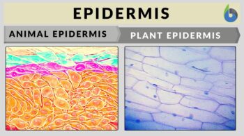
Source: modified by Maria Victoria Gonzaga, from BruceBlaus (human skin diagram, left), CC BY 3.0. Golovetskiy (onion skin, right), CC BY-SA 4.0.
Table of Contents
Epidermis Definition
What is the epidermis? We can define the epidermis as one or more layers of cells forming the tough and protective outer layer of the skin or integument (natural coating). The epidermis is a protective outer covering of many plants and animals. It may be comprised of a single layer, as in plants, or of several layers of cells on top of the dermis, as in those of vertebrate animals.
Human Epidermis
In humans, the skin is the largest organ of the integumentary system. It surrounds the outside surface of the body. The role of the skin is vital as it protects the body (especially the underlying tissues) against pathogens, UV light, chemicals, mechanical injury, and excessive water loss. It is also involved in providing insulation, temperature regulation, and sensation. The layers of skin in humans and other mammals are composed of three major layers: the epidermis, the dermis, and the hypodermis. The primary role of the epidermis is to protect the more susceptible layers of the skin.
Epidermis structure
There are three layers that make up the skin. These include the epidermis, the dermis and the hypodermis. Figure 1 shows the skin structure.
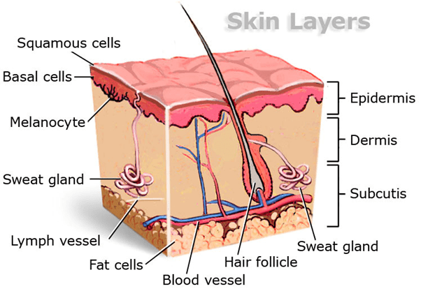
The epidermis is composed of mainly keratinized stratified squamous epithelium. Stratified is defined as being more than one layer thick and squamous refers to the structure of the cells resembling ‘scales’ or having a flattened appearance. The main component of epidermal cells is keratinocytes. These cells produce the protein keratin. Keratin is a strong fibrous structural protein that is a major constituent of outer coverings in vertebrates including skin, feathers, hair, and hooves.
The epidermal cells have no direct blood supply; therefore, they rely on diffusion from blood capillaries in the dermis for oxygen and nutrients.
The epidermis is either made of four or five layers depending on its location. Where there are only four layers, it is described as thin skin, where there are five layers it is described as a thick skin. Skin thickness varies depending on where it is in the body. In general, hairless skin is the thickest because the epidermis has an extra layer known as the stratum lucidum. Thick skin is found on the soles of our feet and the palms of our hands. The layers of the skin will be described in more detail below.
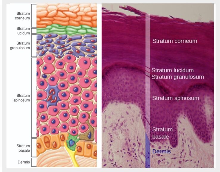
There are five layers of the epidermis. These are the stratum basale, the stratum spinosum, the stratum granulosum, the stratum lucidum, and the stratum corneum. The stratum lucidum is found only in thick skin and is located between the stratum corneum and the stratum granulosum. Figure 2 shows the layers of the epidermis.
(1) The Stratum Basale
The stratum basale, which is also known as the stratum germinativum, is the innermost layer of the epidermis. The cells here are cuboidal or columnar. They lie on the basement membrane that separates the dermis from the epidermis. The stratum basale consists mainly of 2 cell types: (1) keratinocytes and (2) melanocytes. Other cell types, e.g. Merkel cells, are also present in very small quantities.
Most of the cells found in this first layer are keratinocytes that are responsible for replenishing cells lost on the skin surface. These cells divide by mitosis leading to the production of millions of new keratinocytes in the stratum basale every day. The process of mitosis only takes place in this layer of the epidermis as it lies close to the capillaries allowing it to gain plenty of oxygen and nutrients via diffusion. The new cells move the old ones up through the layers of the epidermis. Over time, as these cells move, they alter in their shape and composition. Keratinocytes also help hold the epidermis together.
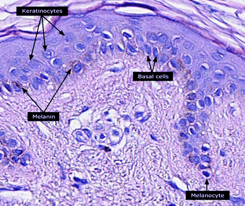
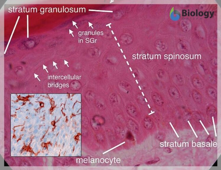
The other main type of cell within the epidermis includes melanocytes which produce melanin. These cells are responsible for giving hair and skin color. Figure 3 shows a sample of keratinocytes and melanocytes stained under a microscope.
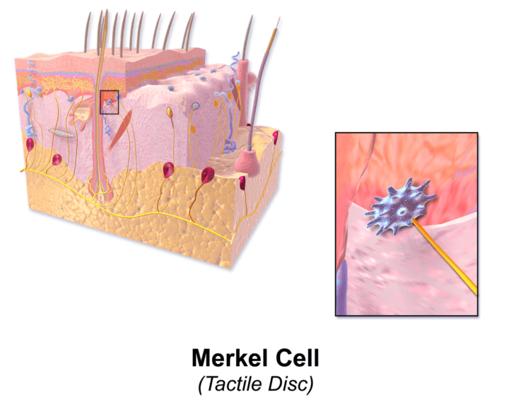
Other cells found in the stratum basale are Merkel cells (Figure 4). Merkel cells were originally discovered by Friedrich Merkel in the late 1800s. They are scarce, specialized epithelial cells found directly above the basement membrane and function as type-1 mechano-receptors. They can sense light/gentle touch as their membrane interacts with nerve endings found in the skin. Therefore, they are found mainly in lips, fingertips, and the face where sensory perception is at its most sensitive. On the other side of their membrane, they are linked to keratinocytes by desmosome multiprotein complexes.
The stratum basale is attached to the basement membrane by desmosomes and hemidesmosomes. These are multiprotein complexes allowing strong cell adhesion. They are intercellular junctions that give mechanical strength to the tissue.
(2) The Stratum Spinosum
The layer above the stratum basale is the stratum spinosum, also known as the prickle layer. It is around 8 – 10 cells thick and it contains polyhedral shaped cells. The cells in this layer contain many desmosomes which function to anchor the cells together. When this cell layer is prepared on a slide for microscopy, the spaces between the desmosomes shrink away giving the cells a spiky appearance. This spiky appearance is not how the cells appear in real life. The desmosomes act as structural support and provide flexibility and elasticity.
Keratinocytes begin to produce keratin in this layer. This strong fibrous protein helps to keep water within the cells. Each keratinocyte contains tonofibrils that begin and end at each desmosome. Tonofibrils are keratin intermediate filaments, components of the cytoskeleton of keratinocytes. Here, they have a very important structural supporting role.
Dendritic cells of the immune system are also found in the superficial portion of this layer. Dendritic cells are termed Langerhans cells when they are found in the epidermis. They lie very close to keratinocytes in this layer. They also extend their processes to the next layer, the stratum granulosum. These Langerhans cells recognize pathogens and allergens in the epidermal layers and migrate to the lymph node where they can initiate an appropriate immune response.
As new keratinocytes move into this layer, they push older keratinocytes out into the next epidermal layer of the skin, the stratum granulosum.
(3) The Stratum Granulosum
The stratum granulosum is around 3-5 cells thick. In this layer, the cells are slightly flattened to a diamond shape. The keratinocytes here produce large quantities of the proteins keratin and keratohyalin. keratohyalin accumulates in granules called the keratohyalin granules. These granules surround the keratin filaments creating an intracellular matrix.
Lamellar granules (or lamella bodies) are also found in this layer of the epidermis. These are secretory organelles that contain glycolipids, enzymes, and proteins that get secreted to the cell surface. They act as glue allowing cells to remain tightly packed together and coat the cells making them waterproof to prevent water loss. This process also prevents the diffusion of nutrients in and out of the keratinocytes. This, in turn, leads to the death of the superficial stratum granulosum, as seen by the loss of their nuclei. The cells in this layer become flattened and squashed in appearance. (See Figure 5)
(4) The Stratum Lucidum
At around 2 – 3 cells thick, this layer is only present in thick skin. At this point, the keratinocytes have died off due to a lack of nutrients and oxygen. This layer is clear and only found on hairless sites of the body where the skin is thick such as the soles of the feet and the palms of the hands. (See Figure 2)
(5) The Stratum Corneum
Finally, the topmost layer of the epidermis is around 20 – 30 cells thick. (See Figure 2) This layer is made of dead keratinocytes that have no nucleus. It is effectively a keratin layer of shrunken, flattened, cornified cells. They can be termed anucleate squamous cells and this layer is sometimes described as the horny layer. The keratinocytes now are almost devoid of water and filled with keratin. At this point, they are known as corneocytes. These cells slough off into the environment.
The dead keratinocytes secrete defensins, which act as part of the immune system. Defensins are antimicrobial peptides that have been found to act against some viruses, bacteria, and fungi.
The entire cycle of a keratinocyte from its generation to when it is sloughed off can take anywhere between 25 – 45 days. Figure 2 shows a microscope image of the epidermis with the stratum basale to the stratum corneum. Note the flaking of the dead cells in the stratum corneum layer.
In humans and other vertebrates, the epidermis consists of the following layers (strata):
- stratum corneum
- stratum lucidum
- stratum granulosum
- stratum spinosum
- stratum basale (or stratum germinativum)
Epidermis functions
The epidermis has many functions including acting as a water barrier, structural stability, immune defense, body homeostasis, endocrine and exocrine functions, and sensation/touch. Each will be discussed in more detail below.
Water barrier and immune defense
Firstly, the epidermis serves as a barrier not only to water, but to microbes, chemicals, and UV damage. It functions as a water barrier due to the hydrophobic inner surface of the plasma membrane. This surface is composed of cross-linking proteins which also gives the epidermis its structural stability. Furthermore, the outer surface of the plasma membrane is also hydrophobic. When the keratinocytes in the stratum spinosum produce lamella granules, they are exported via exocytosis to the intracellular spaces. These lamella granules are filled with ceramides, phospholipids, and glycosphingolipids, which on secretion, help to form the extracellular lipid-rich membrane. This blocks the movement of water. Moreover, fatty acids are antimicrobial and it is also in these lamella granules that further antimicrobial chemicals such as defensins are released.
Structural integrity
The structure of the skin, with its abundance of the fibrous protein keratin, allows the epidermis to act as a protective covering acting against mechanical stress and injury. It is strong and flexible and shields the tissue layers underneath the epidermis. The appearance of a person’s skin allows us to see their general health and age.
Body homeostasis, endocrine, and exocrine functions
In the stratum spinosum and the stratum granulosum layers of the epidermis, vitamin D is produced. This occurs in keratinocytes with the help of UV light from the sun that converts 7-dehydrocholesterol into vitamin D. A vitamin D receptor and the relevant enzymes are also expressed by keratinocytes allowing vitamin D to be converted into its active form. Stimulation of the vitamin D receptor encourages the generation of keratinocytes in the stratum basale and the differentiation of the keratinocytes as they migrate through the layers of the epidermis.
The epidermis also regulates temperature and water loss. Exocrine functions include sweating and the actions of the sebaceous glands. Sebaceous glands are an apocrine gland (a type of exocrine gland) that secrete oils containing lipids and proteins known as sebum which bud off into the area surrounding a hair follicle via ducts. This oil moves up through the hair shaft and then coats the epidermis protecting from water loss. Sebaceous glands are usually located in hairy parts of the body and are not found on the soles of the feet or the palms of the hands. Figure 6 shows the location of the sebaceous gland in the skin.
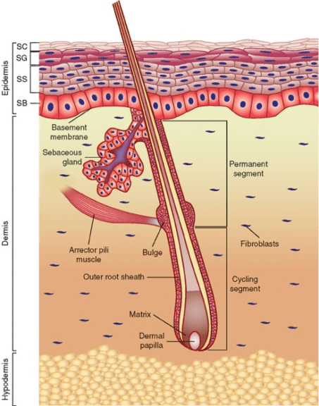
Sweat glands on the other hand are eccrine glands that respond to body temperature. These are found in large quantities in skin tissue throughout the body, particularly the palms of hands and soles of the feet. Their job is to secrete water and electrolytes which disperse through the epidermis allowing the regulation of body temperature through sweating.
Sensation/Touch
As mentioned earlier, Merkel cells are involved in the mediation of the perception of light touch. Merkel cells are described as neuroendocrine cells or mechanoreceptors. They are found in clusters in areas called touch domes. Free nerve endings are also found in the epidermis that aid with sensory functions including sensations to heat, touch, cold, and pain.
More Examples of Epidermis
Below are some of the examples of the epidermis in animal and plant organisms.
Animal epidermis
Keratin in the epidermis of the skin is found in a variety of different animal species. For example, it is a constituent of hooves, nails, claws, scales, feathers, hair, and horns. These appendages are made by modifications of the epidermis as well as the dermis and the subcutaneous layers. Antlers, although composed of bone, have a velvet epidermal covering. (Figure 7)
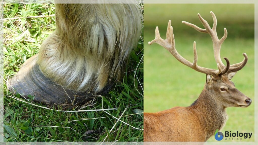
Hooves, nails, claws, and horns are some of the toughest biological materials due to the strength of the dead keratin-filled cells. Due to the complex nature of keratinous material, their formation can serve a wide range of biological properties such as impact resistance (as in hooves), external attack (horns and nails), and withstanding aerodynamic forces (feathers).
Plant epidermis
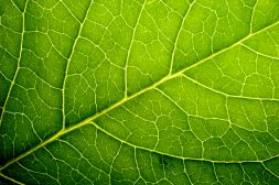
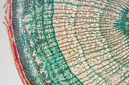
The epidermis of plants is far more simple in structure than the epidermis of animals. Usually, it is only one cell thick, consisting of simple epidermal cells, guard cells, and hairs. However, an epidermis with multiple layers can be found in the leaves of some plants as found in the genus Figus and Nerium. The epidermis in stems contains cells that are tabular in shape. The cells of the epidermis are usually tightly set together with limited intercellular spaces. Figure 8 shows the structure of a leaf indicating the location of the epidermis on the lower and upper sides.
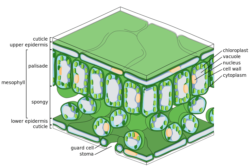
The epidermis covers all herbaceous plants and is found covering the leaves, stems, flowers, and roots. Above ground, the epidermal cells of plants have a waxy coating known as a cuticle which is impermeable to water. (Figure 9) The cuticle keeps water in and pathogens out providing a protective role. An epicuticular wax covers the epidermis of some plants. This further protects plants from water loss as well as wind and strong sunlight. These plants are adapted to living in hot, dry environments. The cuticle is transparent allowing light to go through it. There are little to no chloroplasts in the epidermal layer, these are found in the layers below and as the epidermis is translucent, the sunlight can reach them with ease.

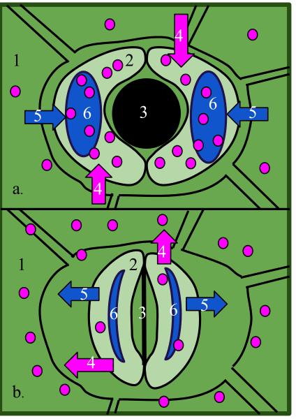
Stomata are present in the epidermis of leaves, these are openings that allow gas exchange. The stomata are made up of a pair of guard cells that make the stomatal pore. These guard cells contain chloroplasts allowing them to make food using photosynthesis. (Figure 10)
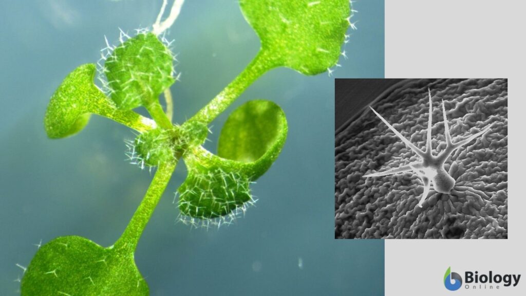
Extensions of the epidermis can be seen in most plants, these are called trichomes that act to protect the plant from insects or herbivores. They can do this by either secreting toxic substances or by preventing pests from reaching the plant surface (spikes or spines). Stinging nettles are one such example where their trichomes can break off and inject the animal/human with histamines that irritate. In insectivorous plants, as in the sundews (Drosera), sticky nectar is produced by the trichomes and this attracts and catches insects for the plant to digest.
The epidermis is also found in the roots of plants. Here, it acts to allow the uptake of water up the root system. The root epidermis has a very thin cuticle to allow water uptake. Roots may produce a chemical called mucigel, which is a carbohydrate that is hydrophilic allowing the root to move through the soil easily.
READ: Plant Tissues – Tutorial
Conclusion
The epidermis is present in animals and plants as an outer protective layer providing a vital barrier to environmental pathogens, chemicals, and UV as well as having an important structural role. In animals, the epidermis has been adapted to individual species to provide protection, defense, and body regulation by the formation of hooves, hairs, feathers, and nails. Similarly, plants have adapted to their environment by either increasing the thickness of their cuticle on the epidermis depending on wet or dry environments. They also have a wide range of trichomes that act to deter pests and herbivores. Overall, the epidermis is accountable for the safety of the organism in which it covers.
Try to answer the quiz below to check what you have learned so far about the epidermis.
References
- Barbieri, J.S., et al. (2014). Skin, Basic Structure and Function. Pathology of Human Disease. 1134-1144. https://doi.org/10.1016/B978-0-12-386456-7.03501-2
- Borradori, L., Sonnenberg, A. (1999). Structure and Function of Hemidesmosomes: More than Simple Adhesion Complexes. Journal of Investigative Dermatology. 112(4) 411-418. https://doi.org/10.1046/j.1523-1747.1999.00546.x
- Cole, A.S., et al. (1988) Epithelium. Biochemistry and Oral Biology. 26 (2) 400-405. https://doi.org/10.1016/B978-0-7236-1751-8.50033-8
- Di Meglio, P., Conrad, C. (2016). Psoriasis, Cutaneous Lupus Erithematosus and Immunobiology of the Skin. Encylopedia of Immunobiology. 5, 192 – 203. https://doi.org/10.1016/B978-0-12-374279-7.15008-8
- Feingold, K, R. (2012). Lamella Bodies: The Key to Cutaneous Barrier Function. Journal of Investigative Dermatology. 132, 1951 – 1953. https://core.ac.uk/download/pdf/82781113.pdf
- Fenner, J., Clark, R.A.F. (2016) Chapter 1. Anatomy, Physiology, Histology, and Immunochemistry of Human Skin. Skin Tissue Engineering and Regenerative Medicine. (1) 1-17. https://doi.org/10.1016/B978-0-12-801654-1.00001-2
- Honari, G., Maibach, H. (2014) Skin Structure and Function. Applied Dermatotoxicology. 1, 1-10. https://doi.org/10.1016/B978-0-12-420130-9.00001-3
- Kobielak, K., et al. (2015). Skin and Skin Appendage Regeneration. Translational Regenerative Medicine. 22, 269-292. https://doi.org/10.1016/B978-0-12-410396-2.00022-0
- Lopez, F.B., and Barclay, G.F. (2017) Plant Anatomy and Physiology. Pharmacognosy. 4, 45-60. https://doi.org/10.1016/B978-0-12-802104-0.00004-4
- The Skin. Lumen Boundless Anatomy and Physiology. https://courses.lumenlearning.com/boundless-ap/chapter/the-skin/
- Sawamura, D., Goto, M., Shibaki, A. et al. (2005) Beta defensin-3 engineered Epidermis Shows Highly Protective Effect for Bacterial Infection. Gene Ther. 12, 857–861. https://doi.org/10.1038/sj.gt.3302472
- Sotiropoulou, P.A. and Blanpain, C. (2020). Development and Homeostasis of the Skin Epidermis. Cold Spring Harbour Perspectives in Biology. https://cshperspectives.cshlp.org/content/4/7/a008383.full
- Wang, B., et al. (2016). Keratin: Structure, Mechanical Properties, Occurrence in Biological Organisms, and Efforts at Bioinspiration. Progress in Materials Science. 76, 229-318. https://doi.org/10.1016/j.pmatsci.2015.06.001
- What is a Merkel Cell? Merkelcell.org. https://merkelcell.org/about-mcc/what-is-a-merkel-cell/#:~:text=Merkel%20cells%20are%20found%20in%20the%20epidermis,outer%20layer%20of%20the%20skin).
- Yousef H, Alhajj M, Sharma S. (2020). Anatomy, Skin (Integument), Epidermis. StatPearls [Internet]. Treasure Island (FL): StatPearls Publishing. https://www.ncbi.nlm.nih.gov/books/NBK470464/
©BiologyOnline.com Content provided and moderated by Biology Online Editors.

