Involuntary muscle
n., plural: involuntary muscles
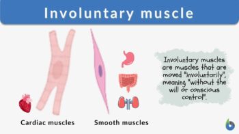
Definition: muscle that contracts without conscious control
Table of Contents
A muscle act typically either under the control of the will or without conscious control. Muscles that can be controlled at will are referred to as voluntary muscles. Those that are not under the control of the will (volition) are called involuntary muscles.
Smooth muscles are involuntary muscles. Find out more here: Smooth muscle vs dense regular connective tissue. Join our Forum now!
Involuntary Muscle Definition
Involuntary muscles are the muscles that contract or move without conscious control. The autonomic nervous system controls involuntary muscle movement. These muscles are generally associated with the viscera or internal organs that exhibit regular, slow contractions and involuntary actions. For example, the heart is an involuntary muscle.
For most involuntary muscles, neuronal stimulation by the autonomic nervous system, hormones, and local factors can trigger the involuntary action of these muscles. However, in certain muscles like the walls of visceral organs, the involuntary muscle contractions can be triggered by the stretching of the muscle.

Involuntary muscle is a muscle that contracts without conscious control. Examples include the smooth and cardiac muscles. The smooth muscles, which are muscles lacking striations when viewed under a microscope. This is why involuntary muscles are sometimes called non-striated or un-striped muscles. The smooth muscles are found lining the internal organs (such as the esophagus, stomach, intestines, etc.) and blood vessels. The cardiac muscle, which is the muscle of the heart, has striations when viewed under the microscope but its contractions are not under the control of the will. The part of the peripheral nervous system associated with the involuntary action of the smooth and cardiac muscles is the autonomic nervous system, which supplies the stimulation to these involuntary muscles. Compare: voluntary muscle
Involuntary Muscles vs. Voluntary Muscles
Functionally, in a human body, the muscles can be classified into two groups:
1. Voluntary muscles
2. Involuntary muscles
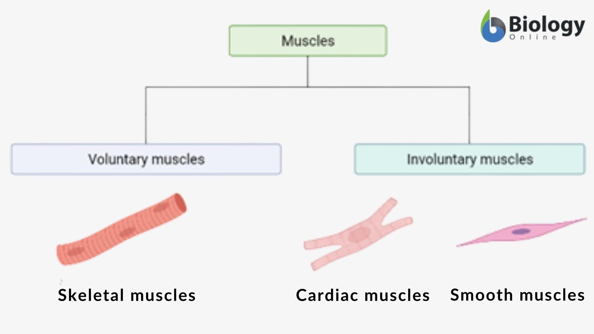
What are involuntary muscles? As suggested by the name, we can define involuntary muscles and voluntary muscles as follows:
Voluntary muscles are those whose movement can be controlled at will or conscious control, while involuntary muscles are those whose movement can not be controlled at will or without conscious control or that work involuntarily, i.e., automatic. Involuntary muscles include smooth muscles and cardiac muscles. This will clear the familiar doubt whether smooth muscles are voluntary or involuntary muscles?
“Cardiac muscle is only found in the heart and needs to have incredible strength and endurance since the heart never stops beating.” Hear more from our Expert: Smooth muscle vs dense regular connective tissue. Join now!
Voluntary muscles (smooth muscle & cardiac muscles) differ from involuntary muscles (skeletal muscle) in several ways; however, the ability to contract involuntarily is of prime importance. Therefore, it is pertinent to understand the difference between these two muscles, enlisted in Table 1.
| Table 1: Comparison Between Voluntary and Involuntary Muscles | ||
|---|---|---|
| Features | Voluntary Muscles | Involuntary Muscles |
| Definition | Definition of voluntary muscle: Voluntary muscles are the ones that move or contract under the conscious control of a person. | Definition of involuntary muscle: Involuntary muscles are the ones that do not move or contract under the conscious control of a person, i.e., these muscles work automatically. |
| Biological system closely associated with | Generally, they are a part of the skeletal system | Generally, they are associated with viscera or internal organs that repeatedly and slowly contract, such as the digestive system and the respiratory system |
| Common names | Voluntary muscles are also known as skeletal muscles or striated muscles | Also known as smooth muscles or nonstriated muscles (although applicable only to smooth muscle types and not the cardiac muscle type) |
| Location | They are located in the skeletal system attached to bones with the help of tendons | These muscles line the organs like the urinary bladder, blood vessels, stomach, intestine, etc. As for the cardiac muscles, they are located in the heart. |
| Muscle cell striations | Voluntary muscles exhibit striations. Striations are the visible bands in the myocytes (muscle cells) of the voluntary muscles that occur due to the organization of myofibrils | Many involuntary muscles do not have striations and they appear to be smooth, as in the smooth muscles. The cardiac muscles have striations. |
| Muscle cell structure | Structurally, the voluntary muscles are unbranched, long cylindrical with the peripherally located nucleus. These muscles may be multinucleated and are rich in mitochondria. | Structurally, the smooth involuntary muscles are long, thin, spindle-shaped cells with a centrally located nucleus. The smooth muscles are uninucleated with a fewer number of mitochondria. |
| Sarcolemma | The voluntary muscle fiber is surrounded by thick sarcolemma. | The smooth involuntary muscle fiber is surrounded by thin sarcolemma. |
| Sarcomere | Sarcomeres are found in voluntary muscle fiber. | Sarcomeres are absent in smooth involuntary muscle fiber whereas present in cardiac involuntary muscle fiber |
| Intercalated disc | Complete absence of Intercalated discs. | Presence of intercalated discs in cardiac involuntary muscle fibers. |
| Nervous system in control | Under control of the somatic nervous system | Under control of the autonomic nervous system |
| Non-myogenic vs. Myogenic | Voluntary muscles are non-myogenic, i.e., the nerve stimuli in such muscles are generated outside by the nervous system. | Involuntary muscles are myogenic, i.e., the nerve stimuli in such muscles are generated in the muscle fiber itself. |
| Movement | Voluntary muscles exhibit rapid and robust movement. | Involuntary muscles exhibit slow and rhythmic movement. |
| Energy requirement | High energy requirement | Smooth involuntary muscle has relatively low energy requirement whereas cardiac involuntary muscle requires high energy |
| Fatigue | These muscles get fatigued and need rest | These muscles do not experience fatigue and work without stopping. |
| Importance | These muscles are essential for all body movement and its locomotion. | These muscles are essential for the functioning of the internal organs and basic functions of life, e.g., heartbeat, elimination of waste products from the body. |
| Examples | Pectoral muscles, hamstrings, biceps, triceps, quadriceps, abdominals, etc. are some of the examples of voluntary muscles. | Cardiac muscle and smooth muscle that line the internal organs like the intestinal tract, blood vessels, urogenital tract, respiratory tract, etc. are involuntary muscles. |
Examples of Involuntary muscles
Below is the list of involuntary muscles. The cardiac muscles and the smooth muscles are types of involuntary muscles found in the body of higher forms of animals, including humans.
Cardiac involuntary muscle
Cardiac muscles are the involuntary muscles that are striated. These muscles are found on the wall of the heart and contract and relax at regular intervals. Individual heart muscle cells are known as cardiomyocytes. Cardiomyocytes are joined together by intercalated discs forming cardiac muscles. Cardiac muscle cells are enclosed in collagen fibers.

Cardiac muscle cells are organized in a parallel fashion and are connected via intercalated discs. These intercalated discs (or glossy stripes) are organized in a Z-shaped or stair-step pattern. Interestingly, cardiac muscles have some elements similar to skeletal muscles, e.g., the myofibrillar structure of sarcomeres.
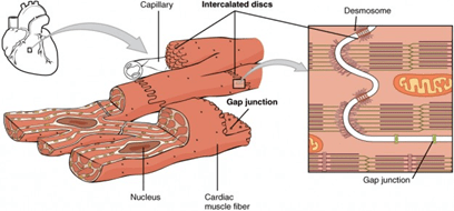
Being myogenic, cardiac muscles differ from skeletal and smooth muscles, and stimulus for contraction is generated within the cardiac muscles. Electrical stimulation generates the action potential in the cardiac muscles for the contraction. As a result of the action potential generation, calcium ions are released into the sarcoplasm reticulum. Elevated levels of calcium ion result in excitation and contraction of the cardiac muscles. Vagal and sympathetic nerves innervate the cardiac muscles and control them.
Smooth involuntary muscle
Smooth muscles are the nonstriated involuntary muscles that line the viscera or the internal hollow organs like, urinary tract, blood vessels, and intestinal tract. The ciliary muscle present in the eye is an example of smooth muscle. Ciliary muscles dilate and regulate the movement of the iris.
Structurally, smooth muscles are fusiform in shape, i.e., round at the center and tapering ends. Smooth muscles are made up of thick and thin filaments that are not arranged into sarcomeres resulting in a nonstriated pattern. Microscopically they appear to be homogenous and hence, named smooth muscles. The cytoplasm of the smooth muscles contains actin and myosin in large amounts. Smooth muscles also have calcium-containing sarcoplasmic reticulum. This calcium-containing sarcoplasmic reticulum is responsible for prolonged contraction.
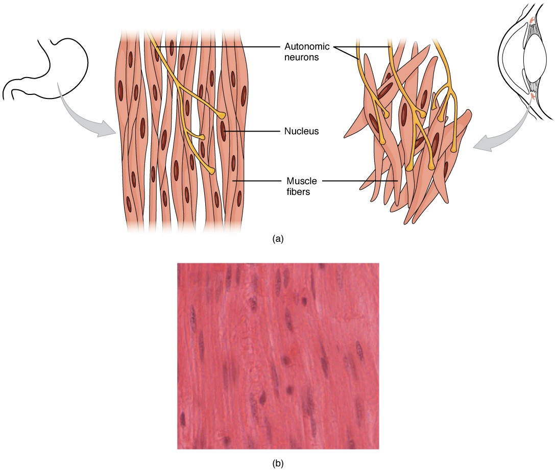
Smooth muscles can be single-unit or multi-unit muscles. Single unit smooth muscles contract and relax as a single unit (i.e., as a whole), while multiunit muscles contract and relax separately. Essentially, multiunit smooth muscles are not electrically coupled and hence show independent contraction/relaxation action. The contractions in the digestive tract are an example of the slow and steady involuntary contractions in the single-unit smooth muscles. On the other hand, smooth muscles lining the lungs’ airways, ciliary muscles in the eye, and the arrector pili muscles in the skin are examples of multiunit smooth muscles.
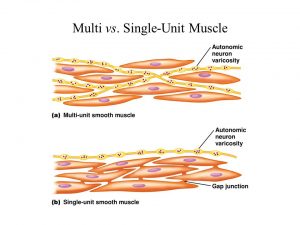
Vesicles of a nerve fiber (or buttons) surround the smooth muscle fibers and carry neurotransmitters. Smooth muscles have a greater elastic property in comparison to striated muscles. The elasticity of smooth muscles is a critical property as it helps to maintain contractile tone in organs like the urinary bladder.
Due to the absence of the sarcomeres, the organization and stretchability of the smooth muscles are not limited. Instead, smooth muscles exhibit a stress-relaxation response wherein the muscles of a hollow organ are stretched when the organ fills up. This mechanical stress due to the stretching of the organ triggers the contraction. However, muscle relaxation immediately after contraction ensures that the organ does not empty its content prematurely. This phenomenon is significant for the urinary bladder, wherein the smooth muscle tone ensures the efficient functioning of the excretory system.
Do you have a question on smooth muscle tissues? Ask our community. Join our Forum: Smooth muscle vs dense regular connective tissue. Let’s talk!
Difference Between Cardiac muscles and Smooth muscles
Though both cardiac muscles and smooth muscles are involuntary, they differ from each other. The differences are:
- Cardiac muscles are striated involuntary muscle tissue found in the heart, while smooth muscles are nonstriated involuntary muscles found generally in the viscera.
- Cardiac muscles are found only in the heart and aorta, while smooth muscles are found in most hollow internal organs and blood vessels.
- The cardiac muscle is innervated by the autonomic nervous system through the cardiac pacemaker, while ANS directly innervates smooth muscles.
- Cardiac muscles never regenerate when injured, while the smooth muscles can.
The Function of Involuntary Muscles
The movement of the involuntary muscles, i.e., involuntary contraction and relaxation, is responsible for basic life functioning. Almost all the hollow organs like the stomach, bladder, and tubular structures like blood vessels, bile ducts, sphincters, uterus, eye, etc., are lined by the smooth muscles, and functions of muscles include the following:
- In the skin, involuntary muscles or smooth muscle cells present in the arrector pili are responsible for the erection of hairs (goosebumps) in response to cold temperature or fear.
- In the eye, ciliary muscles (smooth muscles) are present, cause the dilation and contraction of the iris that eventually changes the shape of the ophthalmic lens.
- Contraction of the vascular smooth muscles present in the arteries and arterioles is responsible for blood pressure and continuous blood flow.
- The involuntary contraction and relaxation of these muscles result in the sealing or closure of orifices (e.g. pylorus, uterine os)
- Contraction of the involuntary muscles or smooth muscles of the digestive tract is responsible for the peristaltic movement in the intestinal tract, resulting in the mixing of food and its movement throughout the digestive tract.
- Similarly, smooth muscle movement in the urinary bladder and urinary tract is responsible for holding up the fluid and its eventual removal from the bladder.
- Involuntary muscles in the heart or cardiac muscles carry out the pumping of blood throughout the body.
- The involuntary movement of the muscles also plays a vital role in the ducts of the exocrine glands.
Try to answer the quiz below to check what you have learned so far about involuntary muscles.
Further Reading
References
- Campbell NJ, Maani CV. Histology, Muscle. [Updated 2021 May 10]. In: StatPearls [Internet]. Treasure Island (FL): StatPearls Publishing; 2021 Jan-. Available from: https://www.ncbi.nlm.nih.gov/books/NBK537195/
- Hafen BB, Shook M, Burns B. Anatomy, Smooth Muscle. [Updated 2020 Aug 15]. In: StatPearls [Internet]. Treasure Island (FL): StatPearls Publishing; 2021 Jan-. Available from: https://www.ncbi.nlm.nih.gov/books/NBK532857/
- Noto RE, Leavitt L, Edens MA. Physiology, Muscle. [Updated 2021 May 12]. In: StatPearls [Internet]. Treasure Island (FL): StatPearls Publishing; 2021 Jan-. Available from: https://www.ncbi.nlm.nih.gov/books/NBK532258/
©BiologyOnline.com. Content provided and moderated by Biology Online Editors.

