Meiosis
n., plural: meioses
[maɪˈəʊsɪs]
Definition: a specialized form of cell division that ultimately gives rise to non-identical sex cells
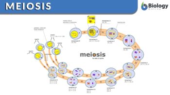
Image source: Modified by Maria Victoria Gonzaga, BiologyOnline.com, from the works of Marek Kultys (schematic diagram of meiosis), CC BY-SA 3.0.
Table of Contents
What is Meiosis?
A simple definition of meiosis would be is this: meiosis is the process of cell division that results in the production of a haploid “daughter” cell with a haploid chromosomal number of a diploid “parent” (“original”) cell. The resulting haploid cell after meiosis would have only one part of the various homologous chromosome pairs of the parent cell.
What are homologus chromosomes, homologues, and sister chromatids? Find the answer here: Difference Between Homologous Chromosomes and Sister Chromatids. Join in now!
Meiosis is a form of cell division in sexually reproducing organisms wherein two consecutive nuclear divisions (meiosis I and meiosis II) occur without the chromosomal replication in between, leading to the production of four haploid gametes, each containing one of every pair of homologous chromosomes (that is, with the maternal and paternal chromosomes being distributed randomly between the cells). Etymology: from Greek meiōsis, meioun (to diminish), from meiōn (less).
The main function of the meiotic division is the production of gametes (egg cells or sperm cells) or spores. In the human body, the meiosis process takes place to decrease the number of chromosomes in a normal cell which is 46 chromosomes to 23 chromosomes in eggs and sperms. So the number of chromosomes in meiosis decreases to half. Consequently, during fertilization when the two haploid cells fuse, the number of chromosomes in the produced cell is restored as somatic cells (each with 46 chromosomes).
The process of meiosis is divided into 2 parts, meiosis 1 and 2. Each part consists of 4 phases (prophase, metaphase, anaphase, and telophase), which is similar to mitosis by being comprised of four phases.
The first part of meiosis (i.e. meiosis I) is the most complicated part of the meiotic division. Prophase I, in particular, occupies almost more than half the time taken for meiosis as it contains 5 substages: leptotene, zygotene, pachytene, diplotene, and diakinesis. The behavior and organization of the chromosomes differ in each stage, which gives clues about the complexity of prophase I.
Meiosis I can be distinguished from mitosis by three main features:
- Meiosis I has reciprocal recombination (may also be called chiasma formation and crossing over)
- Meiosis I has the pairing of the homologous chromosome
- The release of the cohesion sister chromatids in a two-step process occurs in Meiosis I.
These features allow the homologous segregation on the mitotic spindle. Then, the two sister chromatids separate during meiosis II. These various behaviors of the chromosome are described below for the distinctive events happening in each meiosis stage. Moreover, it should be noted that these events are interdependent. This means that the different events during the pairing of chromosomes, such as the recombination of reciprocal, the crossing-over, and the formation of chiasma are connected; therefore, the only successful process of recombination at meiosis I prophase will be the one that produces the correct homologous chromosome segregation at meiosis I.
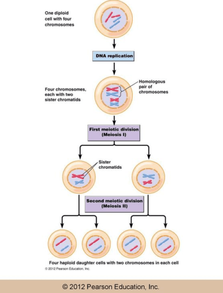
The two succeeding chromosomal divisions result in the halving of the original number of chromosomes. After the completion of S phase and the production of identical chromatids from the replication of the parent chromosome, meiosis I commence. The chromosomes start to pair with each other and eventually segregate into two cells. The chromatids, though, remain together so each of the newly formed daughter cells will contain one of the homologous chromosomes with two chromatids by the end of meiosis I.
Meiosis II follows Meiosis I. The two chromatids will then separate and segregate to two daughter cells. Therefore, at the end of meiosis II, four daughter haploid cells are produced, each containing one copy of each chromosome.
After the replication of DNA, the pairing of the homologous chromosomes does not only allow for the segregation of meiotic chromosomes but also contributes to the recombination of maternal and paternal chromosomes. This pairing of chromosomes occurs during the prophase of meiosis I.
Function of Meiosis
Why is meiosis important for organisms? Imagine this, if gametes (eggs and sperms) were to be produced by mitotic division only and not be meiosis, then the gametes would contain the same number of chromosomes as that of the diploid somatic cells. Consequently, when the gametes fuse during fertilization, the resulting zygote will contain four sets of the homologous chromosome and become tetraploid.
This scenario of “doubled chromosome content” will go on to the next generations and this leads to chromosomal aberrations. The chromosomal number is disrupted and unkept throughout generations. This is, in fact, a case of chromosomal abnormality. Therefore, to keep the number of chromosomes constant in each generation, gametes are produced by the process of meiosis, during the formation of gametes, meiotic cell division decreases the number of chromosomes to haploid.
So what does meiosis produce? Meiosis starts with one round of replication of chromosomal DNA, then two steps of nuclear division. As a result, four daughter nuclei (each of them is present in a new daughter cell) are produced from the meiotic division of the original cell. Each daughter cell nucleus contains only a haploid number of chromosomes. The formation of gametes haploid cells occurs in two rounds: Meiosis I and II, with DNA replication for one time only (at the S phase of interphase).
How do you know if a chromosome is homologous? Our Expert shares insights: Difference Between Homologous Chromosomes and Sister Chromatids. Join our Forum now!
Meiosis vs. Mitosis
What is the difference between meiosis and mitosis? Meiosis and mitosis are the two main forms of cell division. The differences between them are summarized in Table 1.
| Table 1: Main differences between meiosis and mitosis | |
|---|---|
| Meiosis | Mitosis |
| Produces haploid cells (n) | Produces diploid cells (2n) |
| Includes two nuclear divisions | Includes one nuclear division |
| The product is a gamete cell | The product is a somatic cell |
| Responsible for sexual reproduction | Responsible for asexual reproduction |
| Crossing over takes place | No crossing over |
| Four cells are produced | Two cells are produced |
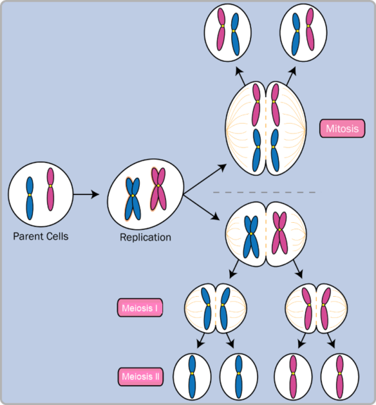
Phases of Meiosis
What is the process of meiosis? Meiosis is the process of four haploid cells formation from a parent diploid cell. The steps of meiosis include 2 stages: meiosis I and meiosis II. Meiosis 1 definition: the first stage in the meiotic division or the reduction division of the meiosis. This is because the number of chromosomes is reduced to half in this stage resulting in the formation of the haploid number of chromosomes.
Each pair of chromosomes come close together to exchange a part of their genetic material in a process or event called a synapse. This process occurs in the early meiosis 1 stages, particularly during prophase I.
During prophase 1 of meiosis I, the homologous pair of chromosomes come very close together and bind tightly to each other so that they almost act as one single unit. This unit is called a bivalent or a tetrad (indicating that each chromosome consists of two sister chromatids so the sum of bivalent is four chromatids). The bivalent splits into two parts after its alignment at the spindle equator so that each chromosome can move to the spindle pole at the opposite side. Consequently, each newly formed daughter nucleus after meiosis I is haploid since it has only one chromosome of the bivalent.
When do sister chromatids separate? Meiosis II which is the second stage of the meiosis cell cycle is somehow similar to mitosis where the two daughter cells are formed as a result of the separation of each two chromatids. Therefore, meiosis I is the stage at which events unique to the meiosis cycle occurs. Nevertheless, each stage of the meiotic division is subdivided in a manner that resembles the mitotic division, such as prophase, metaphase, anaphase, and telophase. However, the prophase of the first meiotic division is much more complicated and longer than the prophase of mitosis. In contrast, the prophase of the second meiotic division is simpler and shorter.
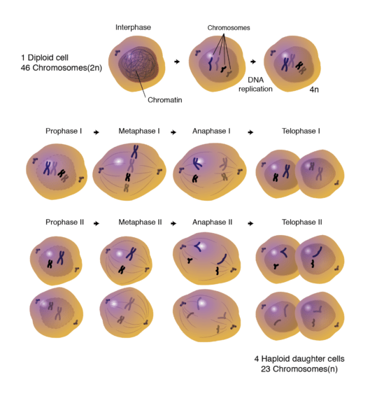
A. Phases of meiosis I
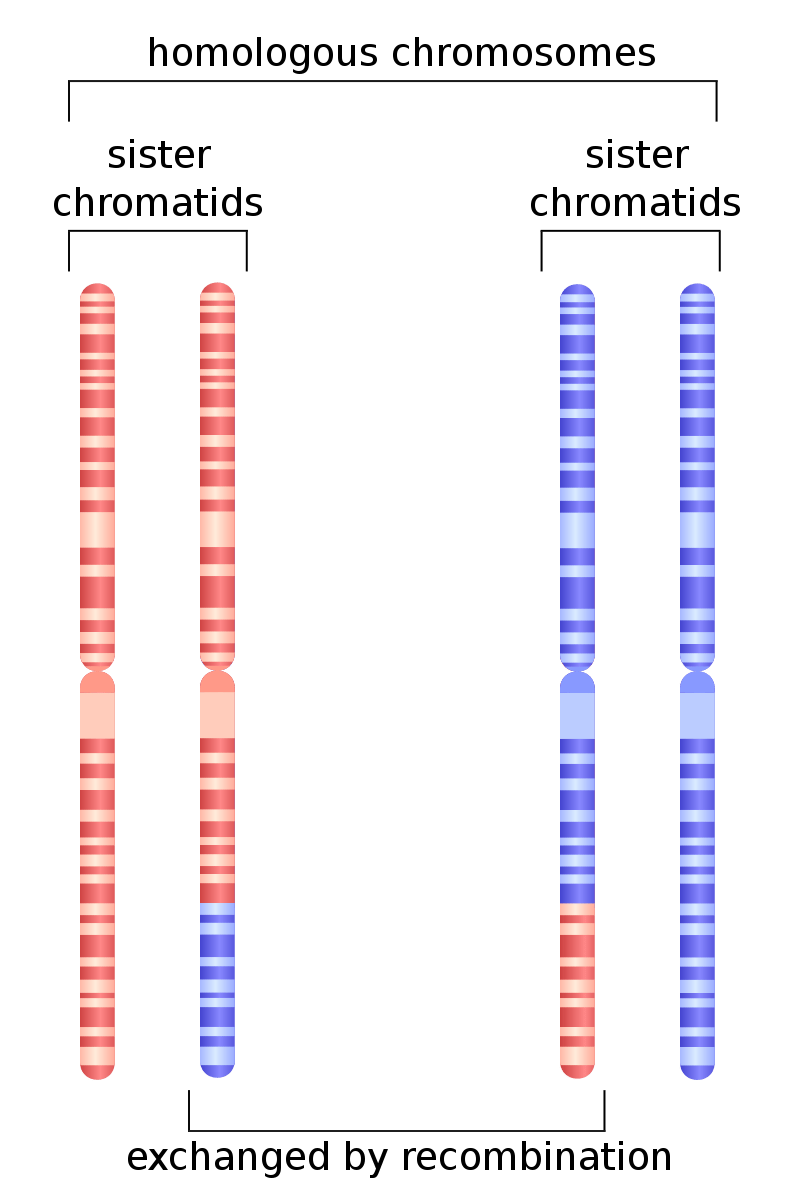
In the cell cycle, meiosis I takes place after interphase where the chromosomes replicate at S phase. Next, the chromosomes condense during the early stages of prophase I. Two centrosomes travel to the two opposite poles of the cell preparing it for nuclear division. Homologous chromosomes consist of pairs of chromatids. These chromosomes form bivalents after pairing in order to be aligned at the spindle equator during metaphase I. Even though homologous chromosomes are separated from each other during anaphase, the two sister chromatids remain attached together.
Step 1: Prophase I
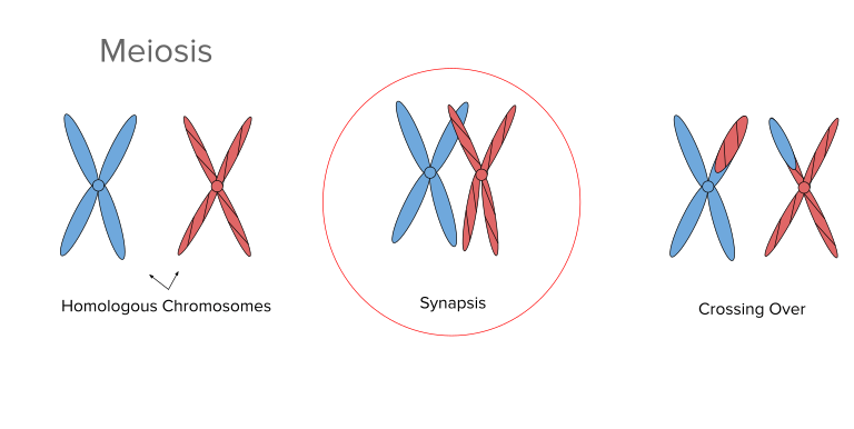
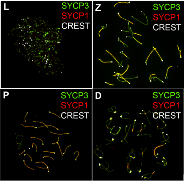
Prophase I is the most complicated phase of meiosis I, it is further subdivided into five stages which are: leptotene, zygotene, pachytene, diplotene, and diakinesis.
The Leptotene stage starts with the chromatin fibers condensing into thread-like-fibers that resemble the formed structure at the beginning of mitosis. The zygotene stage includes further condensation of the fibers that enables them to be distinguished as individual chromosomes. As a result of synapsis, the bivalents ) form when the pairs of chromosomes become tightly paired together. (See figure 4)
The formation of bivalent is critically important in the process of the exchange of the DNA segments containing the genetic material between the two close chromosomes in a process known as crossing over. This process takes place during the pachytene stage. The corresponding segments of chromosomes exchange genetic information for the recombination of genes.
Want more biology facts on homologous chromosome and sister chormatids? Join our Forum: Difference Between Homologous Chromosomes and Sister Chromatids.
Compacting of chromosomes to almost less than a quarter its length occurs during the pachytene stage as well. During the diplotene stage, near the centrosome, the two chromosomes of each bivalent separate from each other. However, the two chromosomes remain attached by chiasmata, which are connections present at the site where the two homologous chromosomes exchange DNA segments.
During diplotene, the transcription resumes, chromosomes decondense, and the cell stops the meiosis for a certain period of time. At the beginning of the final stage of prophase I, the diakinesis, when the chromosomes are re-condensed to their maximum state of compaction, the centrosomes move further.
The chromosomes are only attached by the chiasmata. Here, the spindles form, the nucleoli disappear, and the nuclear envelope disappears. The formation of the meiotic spindle starts and the disintegration of the nucleoli are indications that meiosis prophase 1 ends and meiosis metaphase 1 begins.
Step 2: Metaphase I
During this phase, the bivalents move to the equator of the spindle after attachment to the microtubules using their kinetochores. This process of the bivalent movement to the cell’s equator is typically confined to meiosis I only and does not occur in the mitotic division. There are four chromatids in each bivalent, consequently, each bivalent contains four kinetochores as well. These kinetochores appear close to each other appearing as a single unit facing the same pole of the cell. Such an arrangement allows the attachment of each kinetochore to the microtubules of the spindle pole on the opposite side. This arrangement is the first step that sets for the separation of the chromosomes during the following anaphase. At this stage, the bivalents are randomly arranged, accordingly, the paternal and maternal chromosomes are aligned to one pole of the cell, and therefore, each newly formed daughter cell will receive a mixture of paternal and maternal chromosomes during their movement to the opposite poles during anaphase.
Step3: Anaphase I
The first step in anaphase includes the migration of homologous chromosomes to the spindle poles by the aid of their kinetochore. This step represents one of the main differences between meiosis and mitosis. In mitosis, the sister chromatids separate during mitosis as they are pulled to the opposite poles. In meiosis, the two sister chromatids remain attached together and the homologous chromosomes move toward the spindle poles after separation. This results in the presence of a haploid number of chromosomes in each spindle pole at the end of meiotic anaphase I. The process of chromatid separation during mitosis is mediated by cleaving the two sister chromatids with the aid of an activated enzyme called separase. To stop the action of separase in meiosis, the cell produces a specific protein called shugoshin that prevents the separation of chromatids by protecting the centrosomal site of the chromosome at which the cleavage process takes place.
Step 4: Telophase I
The final phase of meiosis I is telophase 1, which is characterized by the migration of chromosomes to the spindle poles. A nuclear envelope could be formed around chromosomes before cytokinesis to produce two daughter cells of haploid sets of chromosomes. Most of the time, the chromosomes condense after the initiation of meiosis II.
Results of meiosis I
By the end of meiosis I, cytokinesis helps in the production of two cells, each with a haploid nucleus. The chromosomes of each haploid cell will each consist of two chromatids attached at the centromere.
B. Phases of meiosis II
Interphase meiosis begins after the end of meiosis I and before the beginning of meiosis II, this stage is not associated with the replication of DNA since each chromosome already consists of two chromatids that were replicated already before the initiation of meiosis I by the DNA synthesis process. In brief, DNA is replicated before meiosis I start at one time only. The stage of meiosis II or second mitotic division has a purpose similar to that of mitosis where the two new chromatids are oriented in two new daughter cells. Therefore, the second meiotic division is sometimes referred to as separation division of meiotic division.
Step 1: Prophase II
Prophase 2 is the stage that follows meiosis I or interkinesis, it is characterized by the nuclear envelope and nucleolus disintegration as well as the chromatids thickening and shortening in prophase II, and centrosomes replicate and migrate to the polar side. Prophase II is simpler and shorter than prophase I; it somehow resembles the mitotic prophase. On the other hand, prophase II is different from prophase I since crossing over of chromosomes occurs during prophase I only and not prophase II. Metaphase II starts at the end of prophase II.
Step 2: Metaphase II
Metaphase 2 of meiotic division is also similar to metaphase of mitotic division, however, only half the number of chromosomes are present in metaphase II, metaphase II is characterized by the chromosomal alignment in the center of the cell.
Step 3: Anaphase II
It is the stage that comes after metaphase II, in this phase, the sister chromatids separate and move towards the poles of the cell. Anaphase II is similar to mitotic anaphase, where both involve the separation of the chromatids. The kinetochore shortening leads to the movement of sister chromatids to the two ends of the cell.
Step 4: Telophase II
Telophase is the final step of meiosis, during telophase II, four haploid cells are produced from the two cells produced during meiosis I, nuclear membranes of the newly formed cells are fully developed, and the cells are completely separated at the end of this phase. However, during spermatogenesis in humans and other animals, the sperms are not fully functioning at the end of telophase II since they need to develop flagella in order to function properly.
Results of meiosis II
Four haploid cells are produced after telophase II and cytokinesis, each daughter cell contains only one chromosome of the two homologous pairs. The produced haploid cells contain a mixture of genetic information from the maternal and paternal chromosomes. These cells contribute to the genetic diversity among individuals of the same species as well as the evolutionary process of organisms.
Examples of Meiosis
Where does meiosis occur? Meiosis is not restricted to one species, it is included in the life cycle of various organisms such as fungi, plants, algae, animals, and humans.
What is the purpose of meiosis? Meiosis may produce spores or gametes depending on the species where in humans and other animals meiosis produces gametes (sperm cells and egg cells) while in plants and algae meiosis is responsible for the production of spores.
Meiosis in plants and algae
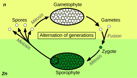
Plants and algae are multicellular organisms that exhibit both haploid and diploid forms of cells in their life cycle. This phenomenon is called alternation of generations where the haploid spores are produced by meiosis. This is also why it is called sporic meiosis in plants and algae. The formed spores germinate and undergo mitotic division giving rise to a haploid plant or a haploid alga. The gametes are produced by mitotic division from the already existing haploid cells; therefore, the haploid form is called gametophyte. The gametes fuse during fertilization to produce the diploid form of cells. The spores are formed from the diploid form by meiosis. Therefore, the diploid form is called the sporophyte.
Meiosis in fungi
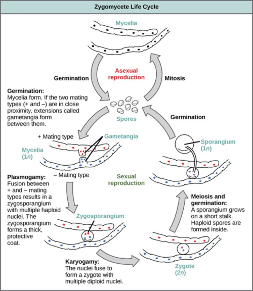
Fungi also have asexual and sexual phases in their life cycle. The mycelium, in particular, may enter either the sexual phase or the asexual phase. When it enters the sexual phase, the haploid mycelia undergoes plasmogamy (the fusion of the two protoplasts) and karyogamy (the fusion of two haploid nuclei). Thus, following karyogamy is the formation of the diploid zygote.
The zygote grows to a stalked sporangium, which by then, will form haploid spores by meiosis. The spores produced by meiosis are called meiospores in contrast to mitospores that are produced via mitosis. These haploid spores (reproductive cells) will be released from the sporangium and each will eventually germinate into a new mycelium. Thus, in fungi, meiosis is the third step in the sequential stages of the sexual phase where plasmogamy is the first followed by karyogamy. Meiosis is crucial in restoring the haploid state of the fungus. See the figure below.
Meiosis in humans and other animals
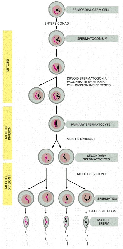
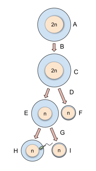
How does meiosis work in humans? Meiosis produces haploid gametes in humans and other animals. It is a crucial part of gametogenesis. As the name implies, gametogenesis is the biological process of creating gametes. In humans and other animals, there are two forms of gametogenesis: spermatogenesis (formation of male gamete, i.e. sperm cell) and oogenesis (formation of the female gamete, i.e. ovum or egg cell).
In oogenesis, four haploid gamete cells are produced from a diploid oocyte. However, only one cell survives and functions as an egg; the other three become polar bodies. This effect results from the unequal division of the oocyte by meiosis where one of the formed cells receives most of the cytoplasm of the parent cell while the other formed cells degenerate which contributes to increasing the concentration of the nutrients in the formed egg. The egg cell acquires most of its specialized functions during phases of meiosis especially prophase I.
In spermatogenesis, the sperm acquires its specialized features in order to develop into a functional gamete after meiosis and post-meiotic events, e.g. spermiogenesis where the sperm cell matures by acquiring a functional flagellum and discarding most of their cytoplasm to form a compacted head.
When does meiosis occur? Meiosis occurs during the reproductive phase of the organism. In humans, though, the meiotic division occurs at different stages. For instance, in males, it starts at puberty and persists throughout their lifetime. In females, the newborn will already have primary oocytes arrested at prophase I and will continue the next stages of meiosis at puberty. However, as each primary oocyte develops into a secondary oocyte at ovulation, it will stop again at metaphase II of meiosis II. Meiosis will only proceed and reach completion at fertilization. If not fertilized, meiosis will no longer proceed and the arrested secondary oocyte will disintegrate. Soon, menstruation begins.
Errors in Meiosis
Meiosis is prone to errors., and therefore, can affect the ability of the human to reproduce. Abnormal meiosis has a great negative impact on human perpetuity. Errors in meiosis steps can result in infertility as well as the formation of gametes of genetically imbalanced features. Meiotic errors are the main contributors to the congenital abnormalities resulting from genetic impairment as well as the mental abnormalities affecting newborn children.
Errors in the pairing and recombination of chromosomes are present in more than 30% of the human oocyte pachytene where the pairing of homologous chromosomes fails, in a phenomenon known as asynapsis.
In yeast, failure in the chromosomal pairing can lead to cell death after triggering the checkpoints of the cell. The same phenomenon is observed in the germ cells of humans. Consequently, the increase in the oocytes with errors in the chromosomal pairing will lead to the depletion in the number of germ cells that result in premature menopause in women. Similarly, errors in the stages of meiosis of spermatocyte production lead to infertility due to the decrease in the number of functional sperms produced.
Depletion in the number of germ cells is more significant in females than in males since the male produces about 300-400 million sperms daily whereas women produce about 300-400 oocytes during her lifetime.
At metaphase I, chromosome pairs might fail to cross over properly, therefore, the unpaired chromosomes segregate randomly with an increased risk of the production of aneuploid gamete, which contains an imbalanced number of chromosomes copies. Moreover, spermatocytes may be eliminated by apoptosis or necrosis due to failed crossing-over.
Biological Importance of Meiosis
What is the function of meiosis in reproduction? For every organ that reproduces sexually, meiosis and mitosis are two essential parts of their cell cycle because of the balance between the number of chromosomes that are doubled during fertilization and the halving of chromosomes during gamete formation by meiosis is maintained. A sexually reproducing organism has a cell cycle that consists of two main phases: a haploid phase and a diploid phase. Meiosis definition biology is the haploid phase that starts during gamete formation and ends with the formation of zygote during fertilization where the diploid phase starts at the formation of a zygote by the fusion of two gametes and ends by meiotic cell division during gamete formation.
How many cells are produced in meiosis? The meiotic division produces four haploid cells from one diploid cell to complete the life cycle of sexually reproduced organisms such as humans and animals. Before meiosis, in the parent diploid cell, the chromosomal DNA duplicates, moreover, four haploid nuclei are formed as a result of two successive divisions of a diploid nucleus.
Why is meiosis important for organisms? Meiosis is biologically important since it is responsible for the genetic diversity among sexually reproduced organisms where during prophase I, the chromatids of the two homologous chromosomes synapse and exchange parts of their genetic materials.
Try to answer the quiz below to check what you have learned so far about meiosis.
References
- Becker, W. M., Kleinsmith, L. J., Hardin, J., & Bertoni, G. P. (2004). The world of the cell (Vol. 4). Menlo Park, CA: Benjamin/Cummings.
- Hultén, M. A. (2010). Meiosis. Encyclopedia of Life Sciences.
- Cooper, G. M., & Hausman, R. E. (2000). A molecular approach. The Cell. 2nd ed. Sunderland, MA: Sinauer Associates.
- Alberts, B., Johnson, A., Lewis, J., Raff, M., Roberts, K., & Walter, P. (2002). Meiosis. In Molecular Biology of the Cell. 4th edition. Garland Science.
©BiologyOnline.com Content provided and moderated by Biology Online Editors.


