Meiotic spindle
n., plural: meiotic spindles
maɪˈɑɪətɪk ˈspɪndəl
Definition: Microtubule-based structure facilitating chromosome segregation
Table of Contents
Meiotic Spindle Definition
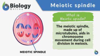
The meiotic spindle refers to the spindle apparatus that forms during meiosis in contrast to the spindle apparatus that forms during mitosis. The spindle apparatus, in turn, is the collective term for all the spindle fibers during cell division — whether mitosis or meiosis. The spindle fibers are responsible for moving and segregating the chromosomes within the dividing cell. Meiosis is a specialized form of cell division that ultimately gives rise to non-identical haploid sex cells. There are two meiotic divisions and each consists of substages: prophase, metaphase, anaphase, and telophase. Depending on the meiotic division, they may be designated as I or II.
Here is an overview of the stages of meiosis characterized in terms of the spindle apparatus:
- The meiotic spindle first forms during prophase I.
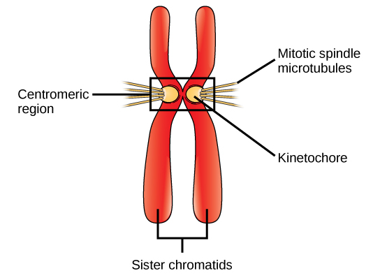
- In the next stage, metaphase I, the spindle fibers attach to the kinetochores of the homologous chromosomes aligned along the metaphase plate.
- In anaphase I, the homologous chromosomes separate as they move apart towards the opposite poles. This movement is through the action of the spindle fibers.
- In telophase I, the spindle fibers disappear.
At this point, the first meiotic division (meiosis I) is over. The original cell has given rise to two cells that will undergo the second meiotic division (meiosis II).
- The meiotic spindle reappears at prophase II.
- In metaphase II, the kinetochore microtubules attach to the kinetochores of the sister chromatids of each chromosome that line up along the metaphase plate.
- In anaphase II, the spindle fibers pull the sister chromatids apart and move them towards the opposite poles.
- In telophase II, the meiotic spindle disappears.
At this point, the second meiotic division (meiosis II) is over. Four haploid cells are produced and will then undergo cellular differentiation to become fully mature gamete.
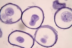
Meiotic spindle (by Francois Nedelec):
Biology definition:
A meiotic spindle is a cellular structure consisting of microtubules emanating from two centrosomes. This spindle apparatus forms during meiosis at opposite poles of the cell. These microtubules attach to and interact with chromosomes, ensuring their proper alignment and segregation during cell division. Etymology: The term “meiotic spindle” originates from the combination of “meiotic,” referring to the type of cell division involved, and “spindle,” denoting its spindle-like appearance under microscopy. Synonym: chromosomal spindle; meiosis spindle. See also: meiosis
Meiotic spindle vs. mitotic spindle
Both meiotic and mitotic spindles are spindle apparatuses with a crucial role during cell division. But in what way are these two structures different? Let us find out!
Table I: Meiotic Spindle vs Mitotic spindle | ||
|---|---|---|
| Aspects | Meiotic Spindle | Mitotic Spindle |
| Occurrence | Involves germ cells that give rise to gametes | Somatic cells |
| Number of Divisions | Participates in two divisions, Meiosis I and Meiosis II | Involves only one division (Mitosis) |
| Formation | Longer in such a way that it first forms in Prophase I, disassembles in Telophase I, and then re-assembles again in Prophase II, and disassembles in Telophase II | Forms in Prophase, disassembles in Telophase |
| Function | Attaches to chromosomes at the kinetochore, and aids in their movement to ensure their proper orientation within the cell as the cell divides | Attaches to chromosomes at the kinetochore, and aids in their movement to ensure their proper orientation within the cell as the cell divides |
| Chromosome segregation | The meiotic spindle segregates the homologous chromosomes during meiosis I, and then the sister chromatids during meiosis II | The mitotic spindle segregates sister chromatids during mitosis |
| Role | (1) Helps maintain chromosomal integrity of the species (by facilitating the movement and segregation of chromosomes, thus, helps produce haploid gametes crucial in maintaining the species’ chromosomal number throughout generations) (2) Promotes genetic diversity by orienting chromosomal pairs at synapses and crossing over | Vital for the proper distribution of chromosomes to daughter cells, ensuring that each cell contains the same genetic material as the other somatic cells |
| Microtubules | The meiotic spindle is made up of microtubules that exhibit a rather more complex behavior in a way that they tend to undergo rapid polymerization and depolymerization as they are involved in processes specific to meiosis, such as the pairing and recombination of homologous chromosomes (in meiosis I) apart from the alignment and segregation of sister chromatids (during meiosis II) | The mitotic spindle is made up of microtubules that are optimized for the alignment of chromosomes and the subsequent segregation of sister chromatids |
Data Source: Maria Victoria Gonzaga of Biology Online
Meiotic Spindle Structure
The meiotic spindle consists of the following components:
- Microtubules: the structural component of a spindle apparatus. These microtubules extend from the centrosomes and then radiate outwards, forming the spindle apparatus. The microtubules that are directly attached to the kinetochore (located at the centromere of chromosomes) are called kinetochore microtubules. Those that are not directly attached to the kinetochore are called non-kinetochore microtubules. Non-kinetochore microtubules interact with other microtubules, and therefore, aid in the overall organizing and stabilizing of the meiotic spindle.
- Centrosomes: the organelles that serve as the microtubule-organizing centers (MTOCs) located at the opposite poles of a dividing animal cell.
- Microtubule-associated proteins: MAPs, such as kinesins and dyneins, help regulate the stability, function, assembly, elongation, and interaction of the microtubules within the meiotic spindle.

Plants do not have centrosomes. Thus, their meiotic spindle is essentially made up of microtubules, organizing centers analogous to MTOCs at various sites within the cell, and MAPs. Because the plant cell divides via a phragmoplast, the meiotic spindle is also associated in part with its formation. Elements of the meiotic spindle are contributed to phragmoplast assembly. The microtubules from the spindle are reorganized at cytokinesis to serve as the tracks that guide the vesicles where they can deposit new cell wall materials for the developing daughter cells.
Function Of Meiotic Spindle
The meiotic spindle is essential for its crucial role in ensuring the proper organization and distribution of chromosomes during meiosis. Here’s how:
- It brings homologous chromosomes together for genetic exchanges essential for generating genetic diversity.
- It facilitates the attachment and alignment of chromosomes, such as the homologous chromosomes along the spindle equator during metaphase I.
- It aids in the subsequent separation of homologous chromosomes during anaphase I, and then the sister chromatids, in anaphase II.
- It helps to produce haploid gametes essential for chromosomal fidelity in sexually reproducing species.
Failure to carry out these functions properly may lead to chromosomal abnormalities, contributing to aberrational conditions, such as aneuploidy and infertility.
References
- Alberts B, Johnson A, Lewis J, et al. Molecular Biology of the Cell. 6th edition. New York: Garland Science; 2014. Chapter 18, Meiosis; Available from: https://www.ncbi.nlm.nih.gov/books/NBK26885/
- Lodish H, Berk A, Zipursky SL, et al. Molecular Cell Biology. 4th edition. New York: W. H. Freeman; 2000. Section 20.3, Meiosis and the Molecular Basis of Heredity; Available from: https://www.ncbi.nlm.nih.gov/books/NBK21506/
- Ma W, Baillat D, Chi T. Meiotic Spindle Assembly and Function. Frontiers in Cell and Developmental Biology. 2020;8:612678. doi:10.3389/fcell.2020.612678
©BiologyOnline.com. Content provided and moderated by Biology Online Editors.

