Metaphase
n., [ˈmɛtəˌfeɪz]
Definition: The stage of cell division characterized by chromosomes aligning along the metaphase plate (equatorial plane) of the cell
Table of Contents
Metaphase Definition
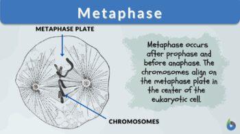
Metaphase is the third phase of mitosis after prophase and before anaphase. Mitosis is the process by which a parent cell’s replicated genetic material is separated into two identical daughter cells. During metaphase, a cell’s chromosomes, which carry genetic information, are aligned at the center (which will eventually split into two sets at anaphase). The chromosomes align on the metaphase plate in the center of the cell during metaphase in eukaryotic cell division. It is the second-most compacted and coiled stage of mitosis. Prophase and prometaphase are the stages that precede metaphase. The chromosomes are condensed, the spindle fibers develop, and the nuclear envelope is broken down during these stages of cell division.
What 3 things happen during metaphase?
The events at metaphase are:
- The chromosomes of a cell compress and migrate together
- Chromosomes align in the dividing cell’s center
- Spindle assembly checkpoint
Process of Metaphase:
The cell goes through a series of checkpoints while it is in early metaphase and late prometaphase to confirm that the spindle has been produced. Microtubules that originate on either side of the cell get attached to the respective chromosomes. During the process of the microtubules retracting, an equal amount of tension is exerted on the chromosomes from both sides of the cell. This moves them to the center of the cell. The process of cell division is finished once the sister chromatids that make up the chromosomes have been separated after the stage of metaphase in the cell cycle.
The centriole divides during the start of eukaryotic cell division and starts forming the microtubule network that will transport chromosomes and organelles throughout the cell division process. These microtubules form bigger fibers that extend from the centrosomes. The fibers are stable around the centrosomes, but they become less stable as they reach out toward the chromosomes. The unstable end of the fibers adds and subtracts components as it comes closer to the chromosome. Fibers wander through the cytoplasm as they expand in this manner, 3 steps forward, 2 steps back. The microtubules can bind to the kinetochore of each centromere.
The checkpoint for the assembly of the spindle is the most essential process that takes place before and throughout the metaphase. The spindle assembly checkpoint is comprised of a complex network of systems that work together to verify that chromosomes are correctly divided.
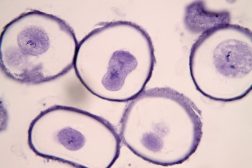
Both mitosis and meiosis go through a spindle assembly checkpoint during metaphase, although the chromosomes align in distinct ways during these two processes. If these checkpoints are missed or do not function properly, the cell will enter anaphase before the chromosomes are correctly connected to microtubules and positioned on the metaphase plate. This might cause errors in the cell’s ability to reproduce. If this is indeed the case, then the chromosomes will be sorted into incorrect cells. This can result in either an excessive number of chromosomes or an inadequate number of chromosomes in the daughter cells that are produced. In the case of meiosis, it can result in birth abnormalities and offspring that are unable to survive. If it takes place at the mitosis chromosome, this can result in cells becoming cancer. The image below is the metaphase microscope and diagram.
 | 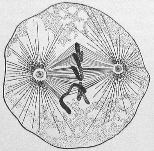 | 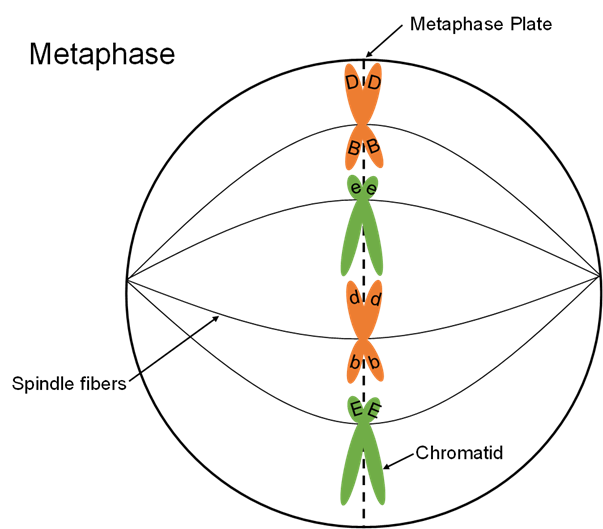 |
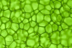
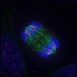
| Metaphase of mitosis. From left, metaphase under a microscope (onion root tip), metaphase in animal cell diagram, and another metaphase diagram with labeled parts. Image Credit (from left to right): Shoba Shanti, Edmund Wilson, and Plant and Soil Sciences. | ||
Watch this vid about metaphase:
Metaphase is the phase following prophase and preceding anaphase of cell divisions (i.e. mitosis and meiosis) and is highlighted by the alignment of condensed chromosomes at the metaphase plate. Cell divisions in eukaryotes, particularly mitosis and meiosis, are important since they give rise to new cells. Mitosis produces two cells that are genetically identical. Meiosis produces four cells that are genetically dissimilar and in which the chromosomes are reduced by half. Both mitosis and meiosis are comprised of these chronological phases: (1) prophase, (2) metaphase, (3) anaphase, and (4) telophase. Since meiosis consists of first and second meiotic divisions, these phases occur twice, each designated as I and II. Metaphase is that phase that follows after prophase when the chromatin condenses and becomes more visible (via the process of chromatin condensation). At metaphase, the condensed chromosomes align at the metaphase plate (cell equator) and the microtubules formed during prophase will attach to the kinetochores. In meiosis, metaphase occurs twice, i.e. metaphase I at first meiotic division and metaphase II at the second meiotic division. Etymology: Greek meta (in the middle) + phase, phásis (“appearance”)”. What is mitosis? What is meiosis?
Metaphase in Mitosis
What is metaphase in mitosis? The process of mitosis metaphase involves the alignment of chromosomes in the center of the cell, with the sister chromatids of each chromosome located on either side of the metaphase plate. During interphase, which occurs between cycles of mitosis, the cell copies its DNA. Before metaphase, the chromosomes that carry the DNA become more compact, ensuring that the DNA is protected from being destroyed by the movements that will take place during metaphase. Chromosomes are dispersed throughout the nucleus in an unorganized fashion during the beginning of metaphase as well as toward the end of prometaphase. Following the dissolution of the nuclear membrane, microtubules will attach to each chromosome.
What moves the chromatids during mitosis? Specialized microtubule structures known as spindle fibers carry out the movement of chromatids during mitosis. During the process of mitosis, microtubules emanating from each centrosome make connections with each chromosome. Cohesins are proteins that join the two sister chromatids that make up each chromosome.
Chromosomes are made up of two sister chromatids. The mitotic metaphase spindle checkpoint needs to be met before the cohesins can be torn apart. This means that all of the chromosomes need to be connected to microtubules from both sides and need to be in the correct position on the metaphase plate. After this checkpoint has been successfully traversed, the chromosomes send out a signal that triggers the anaphase-promoting complex to become active. In the process of mitosis, the activation of this complex brings to the completion of metaphase and the beginning of anaphase.
The fact that the chromosomes are arranged so that sister chromatids are on opposite sides of the metaphase plate assures that the two new cells will be genetically identical. During the synthesis stage of interphase, one chromosome will split into two new strands of DNA, and these two new strands will be known as the sister chromatids. When all of these copies are separated into new cells, they end up with two new cells that are exactly like the original cell from which the cycle started. The mitosis helps to generate new organisms as well as repair damage to existing tissues.
In the meiotic process, the chromosomes rearrange themselves in a new pattern, and the cell divides twice, leading to a reduced genetic material in each daughter cell. Let’s find out more about meiosis below.
Metaphase in Meiosis
Meiosis consists of first and second meiotic divisions. Thus, metaphase occurs as Metaphase I and Metaphase II.
Metaphase I
Prophase, metaphase, anaphase, and telophase are the meiosis stages that occur in both Meiosis I and II. Meiosis I is the first division of the process of meiosis, and during this division homologous chromosomes are separated in a cell. Before the meiosis metaphase begins, the DNA has already duplicated itself, just as it does in mitosis, and all of the chromosomes are in the form of sister chromatids. A homologous pair of each chromosome, share the same segment of DNA but have distinct alleles. There is one homologous pair for each autosome chromosome. The attachment of these homologous pairs to each other occurs during metaphase I in meiosis, as opposed to during mitosis. During metaphase I, the homologous pairs are lined up on the metaphase plate rather than the sister chromatids.
The meiotic spindle checkpoint is the name of the remaining spindle checkpoint that needs to be passed. The cell will be able to move on to anaphase I once all of the chromosomes have been connected to their homologous pair, and once each homologous pair has been attached to microtubules from both sides. During anaphase I, the homologous pairs will be split up into their components. Since of this, the ploidy of the cells will change from diploid to haploid because each new cell will only have a single copy of the genome, and hence only a single allele for each gene. Accidents that occur during metaphase I have the potential to cause cells to have an incorrect number of chromosomes in each cell. Even if only one homologous pair does not separate correctly, the gametes that are produced can result in the production of non-viable progeny. If metaphase I is completed without error, meiosis I will proceed, resulting in the formation of two cells, each of which will have two copies of one complete genome.
Metaphase II
The process of cell division metaphase will start back up again after a brief pause known as interkinesis. As there, is a pause during which there is no replication of DNA, this results in each cell having two copies of a single allele for each gene. During prophase II, the chromosomes undergo another round of condensation, and at the beginning of metaphase II, the nuclear envelope begins to degrade. This time around, on the other hand, there are no homologous pairs; only sister chromatids are present. These chromosomes will become oriented on the metaphase meiosis plate during metaphase II. Once the checkpoint for the meiotic spindle has been reached, the sister chromatids will separate, just as they do during mitosis. After this, the cells can continue to divide until a total of four cells have been generated. These cells will each only have a single copy of each gene’s allele, as well as only a single allele itself.
Metaphase in cytogenetics and cancer studies
The examination of chromosomes in the metaphase state is a fundamental component of both cytogenetics and the study of cancer. Due to the general thickening and the increased coiling of the chromosomes that occur during metaphase, they are at their optimal state for visual examination. The traditional illustration of chromosomes is made up of chromosomes in the metaphase phase (karyotype). For classical cytogenetic analyses, the growth of cells in short-term culture before they are arrested in the metaphase cell state with a mitotic inhibitor. In addition, they are utilized for the fabrication of slides and the banding (staining) of chromosomes so that they can be observed under a microscope, to investigate the structure and quantity of chromosomes (karyotype). The pattern that is produced by staining the slides, most frequently with Giemsa (G banding) or Quinacrine, can include as many as several hundred bands in total. Normal metaphase spreads are put to use in techniques such as FISH, and they also serve as a hybridization matrix for research involving comparative genomic hybridization (CGH).
For the cytogenetic study, malignant cells derived from solid tumors or samples of leukemia can also be employed to make metaphase preparations. Inspection of the stained cell in metaphase chromosomes enables the determination of numerical and structural changes in the genome of the tumor cell. For instance, losses of chromosomal segments or translocations may result in chimeric oncogenes, such as bcr-abl in chronic myelogenous leukemia. These changes can be found by inspecting the stained metaphase chromosomes. Colcemid in metaphase is used during lymphocyte karyotyping for the analysis of chromosomes and also for the analysis of cell chromosomes of amniotic fluid. (Ray, Bhattacharyya, & Biswas, 1984)
Interesting Fact:
Question: Why is the metaphase important?
Answer: The step of metaphase ensures that the daughter cells formed, must be similar to the parent cell. If the step of metaphase gets missed the daughter cells are different from the parent cell and it will cause the formation of the mutated cell.
Answer the quiz below to check what you have learned so far about metaphase.
Further Reading
- Meiosis and Alternation of Generations – Biology Online Tutorial
- Cells know when to separate at mitosis – Biology Online Article
References
- Nature. (2014). metaphase. Retrieved 26 May, 2022, from https://www.nature.com/scitable/definition/metaphase-249/
- NIH. (2022). Metaphase. Retrieved 26 May, 2022, from https://www.genome.gov/genetics-glossary/Metaphase#:~:text=%E2%80%8BMetaphase&text=Normally%2C%20individual%20chromosomes%20are%20spread,when%20viewed%20through%20a%20microscope.
- Ray, K., Bhattacharyya, B., & Biswas, B. B. (1984). Anion‐induced increases in the affinity of colcemid binding to tubulin. European journal of biochemistry, 142(3), 577-581.
- Tutors, V. (2022). Mitosis phases. Retrieved 26 May, 2022, from https://www.varsitytutors.com/high_school_biology-help/understanding-structures-of-mitosis#:~:text=Spindle%20fibers-,Explanation%3A,the%20center%20of%20the%20chromosome.
©BiologyOnline.com. Content provided and moderated by Biology Online Editors.


