Osteon
n., plural: osteons or ostea
[osˈte.ɒn]
Definition: Haversian system
Table of Contents
What is an Osteon?
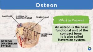
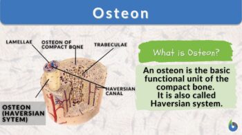
In osteology (the study of bony structures and skeleton), the osteons (or Haversian systems) are the primary structural and functional unit of a compact bone. They are found in the bones of several mammals, birds, reptiles, and amphibians.
Osteons can be several millimeters in length and have a diameter of approximately 0.2 millimeters (0.008 inches). They often run in a direction that is perpendicular to the primary axis of a bone. Osteons are cylindrical structures composed of concentric layers of bone tissue called lamellae that surround a central canal.
The Haversian canal is a long, hollow channel that runs through the center of the osteon, which is the primary unit of compact cortical bone. The osteon is formed of concentric bone layers that are known as lamellae. The Haversian canal contains several smaller blood vessels that are responsible for supplying osteocytes (individual bone cells) with blood.
Osteons are not found in spongy bone (called cancellous bone). Instead, spongy bone is composed of plate or rod-shaped lamellae, which is called trabeculae.
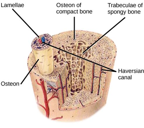
An osteon, also known as a Haversian system, is a cylindrical structure found in compact bone tissue. The osteon provides strength and support to the bone, and it also helps in the repair and remodeling of bone tissue. Osteons can be several millimeters in length and have a diameter of approximately 0.2 millimeters (0.008 inches).
Etymology: from Ancient Greek ὀστέον (ostéon:), meaning “bone”.
Synonym: Haversian system.
Variant: osteone
Watch this vid about osteon:
Structure
Each osteon is made up of several concentric layers (also known as lamellae) of dense bone tissue that surrounds a central canal known as the Haversian canal. The Haversian canal is where the blood supply to the bone is located. The cement line can be thought of as the endpoint of an osteon.
Each Haversian canal is enclosed by a varied number (five to twenty) of lamellae of bone matrix that are arranged in a concentrically spiraling pattern. The lamellae at the surface of compact bone are oriented parallel to the surface; these lamellae are called circumferential lamellae.
Some osteoblasts transform into osteocytes (responsible for mechanical loading), each occupying its own tiny space, or lacuna. The intracellular divisions of neighboring osteocytes are communicated with by a network of tiny transverse canals called canaliculi. This network allows the exchange of metabolic as well as nutritional exchange.
Collagen fibers in one lamellar bone are aligned parallel to one another, whereas collagen fibers in other lamellae are oriented obliquely.
Interstitial lamellae help to maintain the structural integrity of bone tissue, distribute mechanical loads, and may play a role in bone repair and regeneration. They are made up of irregularly arranged collagen fibers and bone cells, and they fill in the spaces between osteons.
The formation of interstitial lamellae is closely linked to the process of bone remodeling.
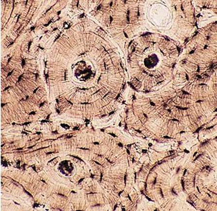
Oblique channels, also known as Volkmann’s canals or perforating canals, connect the osteons to the periosteum. Immature compact bone does not have osteons and has a braided texture. In a remodeling process including bone resorption and new bone formation, the osteons form around a framework of collagen fibers and are eventually replaced by mature bone.
Note it!
Question: What is the function of osteocytes in bone tissue?
Answer: Osteocytes are bone cells that help maintain bone health and integrity by regulating the mineral content and repairing damaged bone tissue.
Drifting osteons
The phenomena of osteon drift are not fully understood. A “drifting osteon” is defined as one that traverses the cortex both transversely and longitudinally. An osteon may “drift” in one or more directions and behind the Haversian canal leaves a tail of lamella.
Drifting osteons can sometimes be observed in bone tissue samples under a microscope, and their presence can indicate ongoing bone remodeling and turnover. However, excessive or abnormal drifting of osteons can be a sign of underlying bone pathology and may require further investigation and treatment.
Serial slices were used in the study to follow drifting osteons longitudinally across the cortex of long bones of baboons and humans. At several cross-sectional levels of the identical systems, the pattern of transverse drift was documented. For each system, the maximum angular variation in the drift direction was measured.
Furthermore, the 3-D model study reveals that the basic multicellular units (BMUs) of drifting osteons are having different morphological characteristics as compared to the BMUs of other osteons of different types. The specific stimuli that activate and guide the drifting BMUs remain uncertain, but it is believed that the complex strain environment experienced by the cortices of long bones may significantly influence their shape. (Robling & Stout, 1999)
Investigative Applications
In forensic investigations and bioarchaeological studies, osteons in a bone fragment are applied to assess an individual’s age and sex, in addition to characteristics of taxonomy, food, health, and motor history. This detail can also be utilized to reconstruct the individual’s motor history.
Since the osteons themselves and how they are arranged vary from taxon to taxon, it is possible to distinguish between genera and even species based on a bone fragment that would not be otherwise identifiable. However, because the properties of the various bones that make up a skeleton can vary quite a little from one another, and because the traits of some faunal osteons overlap with those of human osteons, the examination of osteons is not the most useful technique for the analysis of osteological remains.
Osteohistology can positively influence studies and provide significant information for a range of investigative applications in several fields, including medicine, archaeology, and paleontology. Here are a few examples of how osteons are used:
Forensic Investigations
An individual’s age can be estimated using the presence of osteons in bone tissue as well as the arrangement of those osteons, which can be used by forensic investigators. When bone tissue ages, the number of osteons that are present in a particular location diminishes, while at the same time, the size of the osteons grows. Forensic investigators can determine an individual’s age at the time of their death by dissecting a bone, counting the number and size of the osteons within the bone microstructure, and studying the bone’s cross-section.
Medical imaging
Osteons can be visualized in medical imaging techniques such as X-rays, CT scans, and MRI scans, which can provide valuable information about the structure and health of bones. Osteons can be used to diagnose bone diseases and monitor bone healing after fractures or surgeries.
Medical applications
Osteon research has led to advances in the treatment of bone diseases such as osteoporosis and osteoarthritis. Researchers are investigating the use of osteons to develop new therapies and treatments, including drug delivery systems and tissue engineering.
Paleontological Studies
Paleontologists can establish the lifestyle and activity habits of extinct animals by analyzing the arrangement and density of osteons in the bone tissue of fossilized remains. For instance, animals that were more active and put more stress on their bones would have denser bone tissue with a higher number of osteons per unit area.
In the past few decades, osteohistological research works of dinosaur fossils have been employed to resolve several problems, such as the question of whether or not dinosaurs were warm-blooded, the periodicity of dinosaur growth and whether or not it was uniform across species, and the periodicity of dinosaur growth and whether or not it was uniform across species.
Biomechanics
Osteons are responsible for the mechanical properties of bone tissue, such as strength, stiffness, and toughness. Understanding the structure and organization of osteons can help researchers design materials with similar mechanical properties, such as artificial bone implants.
Biomedical Research
In the field of biomedical research, osteonal bone can be a valuable source of information regarding development and bone regeneration. Researchers can acquire insights into the mechanisms of bone formation, growth, and remodeling, as well as the impact of diseases and injuries on bone tissue, by investigating the structure and arrangement of osteons in bone tissue. This allows the researchers to better understand how bone grows and changes. In the field of biomedical research, osteons have been utilized, particularly, in the process of developing techniques for bone tissue engineering.
Materials science
Osteons are being used as a model for designing synthetic materials with similar mechanical properties. Researchers are exploring how the structure of osteons can be replicated in engineered materials, such as ceramics and composites, to improve their strength and durability.
In summary, the study of osteons has many applications in medicine, archaeology, paleontology, and biomechanics. By understanding the structure and function of osteons, researchers can gain insights into bone health, human history, and material science. (Physiopedia, 2023) (Florencio-Silva, Sasso, Sasso-Cerri, Simões, & Cerri, 2015)
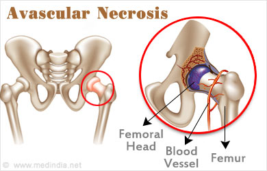
“The Importance of Osteon: the case of femoral neck fractures and bone death”
The femoral neck is a part of the thigh bone (femur) that connects the ball-shaped head of the femur to the shaft of the bone.
The femoral neck is a common site for fractures, especially in older adults, due to the weakened bone density that comes with age.
Fractures of the femoral neck can disrupt the osteons and damage the blood supply, leading to a decrease in bone strength and increased risk of complications, such as avascular necrosis (death of bone tissue due to lack of blood supply) or delayed healing.
Take the Osteon – Biology Quiz!
References
- Britannica, T. E. o. E. (2023). compact bone. Retrieved 12 March, 2023, from https://www.britannica.com/science/fibula-bone
- Florencio-Silva, R., Sasso, G. R. d. S., Sasso-Cerri, E., Simões, M. J., & Cerri, P. S. (2015). Biology of bone tissue: structure, function, and factors that influence bone cells. BioMed research international, 2015.
- Physiopedia. (2023). Functional Unit of Compact Bone. Retrieved 12 March, 2023, from https://www.physio-pedia.com/Bone
- Robling, A. G., & Stout, S. D. (1999). Morphology of the drifting osteon. Cells Tissues Organs, 164(4), 192-204.
©BiologyOnline.com. Content provided and moderated by Biology Online Editors.

