Proteoglycan
n., plural: proteoglycans
[ˌprəʊtɪəʊˈɡlaɪkæn]
Definition: a heavily glycosylated protein
Table of Contents
What are proteoglycans? Proteoglycans are primarily a type of polysaccharide. Structurally, proteoglycans are macromolecules comprised of a protein core covalently attached to glycosaminoglycans. Proteoglycans are found in connective tissues, extracellular matrices, and cell surfaces and function by providing a hydrating, lubricating, and protecting gel structure in and around the cell.
Let’s find out more about proteoglycans starting with the definition of proteoglycans and then moving to their characteristics, types, general structure, function, synthesis, and biological importance.
Proteoglycan Definition
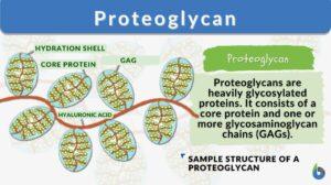
Proteoglycan is a type of glycoprotein characterized by being made up of a protein core covalently attached to one or more glycosaminoglycan chains (GAGs). Proteoglycans produced by the cells may be found on the cell surface (for cell signaling and signal transduction) or exported in secretory vesicles into the extracellular matrix of the tissue where they fill the spaces between cells, forming complexes with other compounds such as collagen, hyaluronan, and other proteoglycans. They are also important in determining the viscoelastic properties of joints and other structures subject to mechanical deformation.
GAGs are the anionic glycan molecules that are made up of monomeric units of N-acetylglucosamine and either glucuronic or iduronic acid. There are primarily six major GAGs in a mammalian cell, namely, chondroitin sulfate, dermatan sulfate, keratan sulfate, heparan sulfate, heparin, and hyaluronic acid (HA).
The number of GAG chains in each proteoglycan can vary from 1 (e.g., decorin) to 100 (e.g., aggrecan). Due to the presence of the GAG chains in their structure, proteoglycans provide a highly hydrated gel that helps to provide cushioning against the compressive forces.
Also, different GAG chains impart different functionality and biological activity to the proteoglycans. Since GAG chains are sulfated as well as non-sulfated (e.g., hyaluronic acid), sulfated proteoglycans and non-sulfated proteoglycans exist.
Proteoglycans are, therefore, macromolecules belonging to a family of heterogeneous polysaccharides. This family has 43 members of different protein cores and GAGs.
Watch this vid about proteoglycans and glycosaminoglycans:
A proteoglycan is a macromolecule that has a core protein with one or more glycosaminoglycan chains. Proteoglycans are a type of glycoproteins present in the body, especially in connective tissues, bone and cartilage, and cell surfaces. Examples of proteoglycans are versican (a large chondroitin sulfate proteoglycan), perlecan, neurocan, aggrecan, brevican, fibromodulin, and lumican.
Where Are Proteoglycans Found?
Proteoglycans are one of the most important and critical polysaccharide components of the extracellular matrix. This remarkably diversified group of polysaccharides is found in the extracellular matrices, connective tissue, and cell surfaces.
The extracellular matrix of a cell is primarily made up of proteoglycans (which are GAG chains attached to proteins) and fibrous matrix proteins, such as collagen. Almost all living mammalian cells form proteoglycans that either become a part of the extracellular matrix or plasma membrane or secretory granules.
Since proteoglycans are an essential part of the extracellular matrix, they are part of the most abundant tissue in the body.
Mast cells are examples of cells rich in proteoglycans. They are characterized by a large content of secretory granules, including mast cell proteoglycans of heparin or chondroitin sulfate proteoglycans of serglycin type. (Rönnberg et al., 2012)
Proteoglycans and Glycoproteins
We know that proteoglycans are glycoproteins but let us understand how proteoglycans differ from glycoproteins. See Table 1.
Table 1: Glycoprotein vs proteoglycan | |
|---|---|
| Glycoprotein | Proteoglycan |
| Define Glycoprotein: Any protein that is attached to a carbohydrate group or chain is referred to as a glycoprotein. | Define Proteoglycan: A core protein attached to glycosaminoglycan chains is referred to as a proteoglycan. |
| Chains attached to the protein core are short, branched with any charge | Long, linear, and negatively charged GAG chains are attached to the protein core |
| Primarily found on the cell membrane as transmembrane proteins | Primarily found in the extracellular matrix or plasma membranes of the cell |
| Involved in the cell recognition and cell signaling | Provides the structure to the cell and is involved in hydration and lubrication of the cell |
| Two types exist based upon protein interactions, i.e., N-linked glycoproteins and O-linked glycoproteins | Classified based upon the present GAG chains |
| Examples: collagen, mucin, etc | Examples: heparin sulfate, chondroitin sulfate, etc. |
Data Source: Dr. Amita Joshi of Biology Online
Note it!
The size of proteoglycan may vary from 10 to 400 kDa.
Characteristics of Proteoglycans
A distinguishing characteristic of the proteoglycan from other glycoproteins is the attachment of the protein core through O-glycosidic bonds to GAG chains.
Proteoglycans are highly glycosylated large protein structures that are either found in the extracellular matrix or attached to the cellular surface.
The majority of the proteoglycans in the extracellular matrix carry out cellular functions/cellular processes as a structural component or as a ligand for several growth factors, cytokines, or chemokines that eventually regulate the inflammatory response, intercellular communication, and embryonic development.
In general, a proteoglycan, with a small size or low-density GAG chains, functions in cell signaling or organization of tissues.
Types
Proteoglycans are composed of core protein and GAGs glycosaminoglycan chains. Proteoglycans are classified either based on the function or based on the GAG chains.
Based on form and function
Accordingly, as per the first classification, proteoglycans have been classified based on their form and function into four major classes:
- Class 1: This class includes proteoglycans that are found and function in the intracellular proteoglycans in secretory granules.
- Class 2: This class of proteoglycans is found and functions on the cell surfaces. These proteoglycans are further classified as
- Transmembrane
- GPI-anchored
- Class 3: These proteoglycans are found and function in the pericellular basement membranes zone
- Class 4: These proteoglycans are found in the extracellular matrix and are further classified as
- Hyalectan-lectincan proteoglycans
- Spock proteoglycans
- Small leucine rich proteoglycans (SLRPs)
Based on GAG component
Proteoglycans can also be classified based on the GAG chains (GAG proteoglycans). Accordingly, chondroitin sulfate-type proteoglycan, dermatan sulfate-type proteoglycan, keratan sulfate-type proteoglycan, heparan sulfate-type proteoglycan, heparin-type proteoglycan, and hyaluronic acid-type proteoglycans are found. The major proteoglycans examples are:
- Aggrecan– This keratin sulfate and chondroitin sulfate type (100-150 GAG chains) proteoglycan is found in cartilage and chondrocytes. Aggrecan functions to provide hydration in the extracellular matrix of cartilage tissues.
- Versican– This chondroitin sulfate type (12-15 GAG chains) proteoglycan is found in smooth muscle cells, mesangial cells of the kidney, brain cells, skin, and fibroblasts.
- Syndecan- Syndecan proteoglycan is a heparin sulfate and chondroitin sulfate type proteoglycan is found in lymphatic cells, mesenchymal cells, plasmocytes, and embryonic epithelium cells. These are transmembrane proteoglycans wherein the extracellular part of the proteoglycan is attached to heparin, collagen fibrils, and fibronectin (matrix fibers) while the intracellular structures are attached to cytoskeletal actin.
- Perlecan- This heparin sulfate type proteoglycan is found in the basal/ basement membrane and is involved in the inflammatory response, angiogenesis (the formation of new blood vessels), wound healing, and bone formation.
- Decorin and Biglycan– This chondroitin sulfate or dermatan sulfate type proteoglycan contain only one chain of GAG and is rich in leucine and is primarily found in fibroblasts, connective tissues, fibroblasts, cartilage, and bones. These proteoglycans primarily function in collagen fibrogenesis.
- Other common proteoglycans include: Heparan sulfate proteoglycans (HSPG) are glycoproteins wherein heparan sulfate chains are present. They are found to be associated with endothelial cells (or endothelium) in vivo. (Key et al., 1992) Cell surface HSPGs are extracellular proteoglycans. Similarly, in chondroitin sulfate proteoglycan, chondroitin sulfate GAG is present. This proteoglycan is involved in modulating cell adhesion, growth, and cell migration. On the other hand, hyaluronic acid proteoglycan is a unique proteoglycan containing a hyaluronic acid GAG chain, which is the only non-sulfated GAG. Keratocan is a specific proteoglycan found in the cornea.
Structure of Proteoglycans
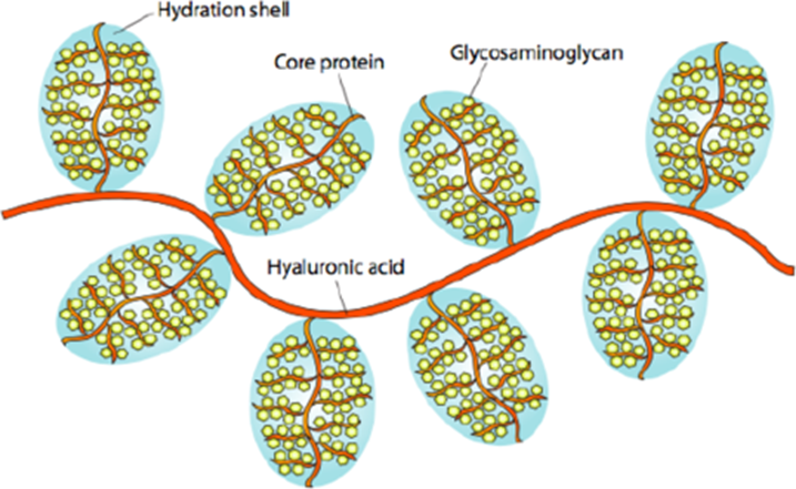
Proteoglycans are highly glycosylated proteins. The basic chemical structure of the proteoglycans is a core protein unit attached to GAG chains (polysaccharides).
Carbohydrate
Almost 95% structure of a proteoglycan is a GAG chain, which is essentially a polysaccharide and hence proteoglycans are more of a polysaccharide than a protein, more precisely proteoglycans are heteropolysaccharides.
GAGs are unbranched polysaccharides made up of repeating disaccharide units of uronic acid (glucuronic acid or iduronic acid) along with an amino sugar (N-acetylglucosamine OR N-acetylgalactosamine).
GAGs are attached to the core protein by glycosidic bonds via tetrasaccharide linker. The linker is made up of:
- (1) a glucuronic acid
- (2&3) two galactose
- (4) a xylose
However, certain linkers may lack glucuronic acid and therefore in such a case trisaccharide linker is present. The GAG chains are attached to the core protein in a perpendicular direction, giving it a brush-bottle-like appearance.
GAG chains are negatively charged and the presence of different GAG chains imparts different functionality to the proteoglycan molecules.
Core proteins
The core protein is the central structure of a proteoglycan molecule to which GAG chains are attached. The core protein may possess amino acid- serine residue in order to connect with the GAG chain. The core proteins or the proteins involved are conserved proteins that are rich in threonine and serine.
Related Stories
The sugars or the carbohydrates attached to the core protein may be carboxylated or sulfated. These carbohydrates or the GAG chains are negatively charged (both types of GAG chains i.e., non-sulfated and sulfate charge are negatively charged chains) and hence, get attracted to positively charged ions that in turn attract more water. This property imparts water gelling property to proteoglycan.
Glycosaminoglycan-substitution domains
The protein core is attached to the GAG chains and these protein cores are rich in serine. The presence of multiple serine residues in the protein core provides the attachment site for multiple GAG chains to the protein core.
In the majority of the proteoglycans, the GAG chains are attached to the protein core via linkers by O-linkages, However, in certain proteoglycans, there can be N-linkages as well. For example, in keratan sulfates proteoglycan, either N- or O-linkages are present.
Keratan sulfates are of two types, I and II. In keratin sulfate I, proteoglycans have N-linkage whereas keratan sulfates II has O-linkages.
The addition of the GAG chain to the serine residue of the protein core is catalyzed by specific glycosyltransferases enzyme. Accordingly, β-xylosyltransferase catalyzes the addition of xylulose while galactosyltransferase adds galactose residues and glucuronosyltransferase adds glucuronic acid residue.
Did you know …?
In human skin, the most abundant proteoglycans are decorin and versican.
Proteoglycan Function
Proteoglycans are multifaceted molecules that serve several biological functions-
- Proteoglycans are part of the extracellular matrix of a cell and are involved in the extracellular matrix function of swelling and hydration that helps the cell to withstand the compression forces.
- Proteoglycan in cartilage forms the cellular structural component of the tissues that help in hydration, lubrication, and protection.
- Proteoglycan serves to promote growth factor sequestering
- Proteoglycans are also involved in inhibiting or inducing angiogenesis or even enhanced angiogenesis
- Proteoglycans are also involved in moderating cell growth, cell proliferation, adhesion, and regulation i.e., proteoglycans play a critical role in cellular growth control
- Cell surface proteoglycan is also involved in intercellular cell signaling pathways
- Proteoglycans are also involved in the release of inflammatory cytokines and chemokines from inflammatory cells
Note it!
Proteoglycans inhibit neural growth and are responsible for the non-regeneration of neural cells.
Synthesis
The synthesis of proteoglycans occurs in ribosomes and is subsequently transported to the lumen of the rough endoplasmic reticulum. Thereafter, after multiple and sequential enzymatic actions by biosynthetic enzymes, glycosylation of proteoglycans occurs in the Golgi bodies. The final proteoglycan is eventually transported to secretory vesicles, which may be released into the extracellular matrix as ECM proteoglycans.
Biological and Clinical Significance
Proteoglycans are the biomolecules that are involved in extracellular matrix modeling, cellular homeostasis, and intercellular cell signaling. Further, proteoglycans, being negatively charged biomolecules, interact with several molecules like growth factors, cytokines, chemokines, etc.
This led to the development of Glycan therapy for the management of various complex diseases like cancer, tissue regeneration, etc. For example:
- Glypican has been found to be a potential target for suppressing metastasis in gastric cancer.
- Syndican has the potential for tissue regeneration and wound healing especially in non-healing wounds.
- Testican has been found to be involved in cancer drug resistance.
- Chondroitin sulfate has been found effective for the management of osteoarthritis.
- Biglycan has been found as a potential candidate for the treatment of Duchenne muscular dystrophy.
Also, ongoing cell biology and pathobiological research point towards the role of proteoglycans in tumor angiogenesis.
Though these proteoglycans have been found to be effective in the management of various diseases, translation of these candidates into therapeutic modalities remains challenging.
Answer the quiz below to check what you have learned so far about proteoglycans.
References
- Couchman, J. R., & Pataki, C. A. (2012). An introduction to proteoglycans and their localization. The journal of histochemistry and cytochemistry : official journal of the Histochemistry Society, 60(12), 885–897. https://doi.org/10.1369/0022155412464638
- Varki, A., Cummings, R., Esko, J., et al., (1999) editors. Essentials of Glycobiology. Cold Spring Harbor (NY): Cold Spring Harbor Laboratory Press; Chapter 11, Proteoglycans and Glycosaminoglycans. Available from: https://www.ncbi.nlm.nih.gov/books/NBK20693/
- Barash, U., Cohen-Kaplan, V., Dowek, I., Sanderson, R. D., Ilan, N., & Vlodavsky, I. (2010). Proteoglycans in health and disease: new concepts for heparanase function in tumor progression and metastasis. The FEBS journal, 277(19), 3890–3903. https://doi.org/10.1111/j.1742-4658.2010.07799.x
- Key, N. S., Platt, J. L., & Vercellotti, G. M. (1992). Vascular endothelial cell proteoglycans are susceptible to cleavage by neutrophils. Arteriosclerosis and Thrombosis: A Journal of Vascular Biology, 12(7), 836–842. https://doi.org/10.1161/01.atv.12.7.836
- Rönnberg, E., Melo, F. R., & Pejler, G. (2012). Mast Cell Proteoglycans. Journal of Histochemistry & Cytochemistry, 60(12), 950–962. https://doi.org/10.1369/0022155412458927
©BiologyOnline.com. Content provided and moderated by Biology Online Editors.

