Pseudopodia
n., singular: pseudopodium
[ˌsjuːdəʊˈpəʊdɪə]
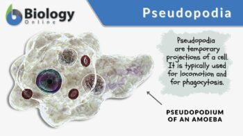
Definition: arm-like, temporary projections of a cell
Table of Contents
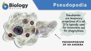
A pseudopodium (plural: pseudopodia) refers to the temporary projection of the cytoplasm of a eukaryotic cell. Pseudopodia are arm-like projections filled with cytoplasm. The projecting cytoplasm, in turn, primarily contains cytoskeleton, such as actin filaments, intermediate filaments, and microtubules. True amoeba (genus Amoeba) and amoeboid (amoeba-like) cells form pseudopodia for locomotion and ingestion of particles. Pseudopodia form when the actin polymerization is activated. The actin filaments that form in the cytoplasm push the cell membrane resulting in the formation of temporary projection. Pseudopodia may be classified into lobopodia, filopodia, reticulopodia, axopodia, and lamellipodia. The most common is lobopodia. Nevertheless, amoeba and amoeboid cells may form in more than one type at once.
Pseudopodia Definition
Biology definition:
Pseudopodia are temporary projections of the cell membrane of eukaryotic cells. And by temporary, it means that it is not a fixed structure. Single-celled organisms characterized by the ability to form arm-like protrusions that can be protracted or retracted are referred to as amoebae. In fact, it is this feature that gave them their name owing to their ability to constantly change their shape. The irregular cell shape is due to their distinctive protoplasmic streaming and their ability to form pseudopodia that deform cell boundaries.
Etymology: The term ‘pseudopodia’ comes from Greek pseudḗs, meaning “false” or “lying” and Greek podós, from poús, meaning “foot” or “leg”. Synonym: pseudopods
Amoeboid Cell Structure
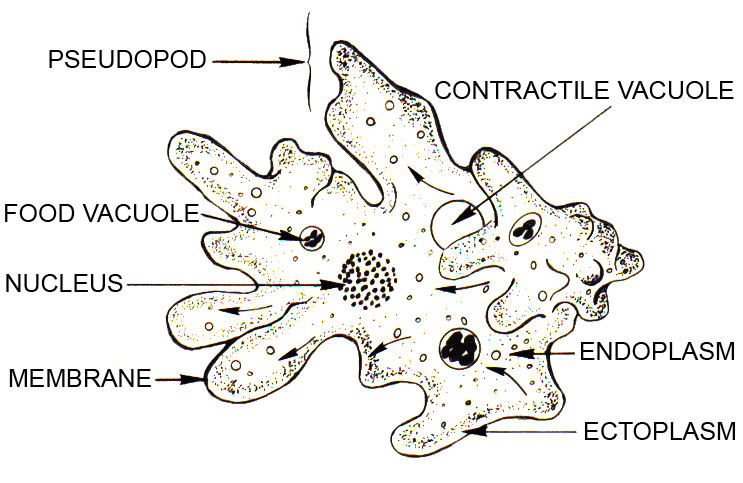
The cell that forms pseudopodia is referred to as amoeba or amoeboid. The term amoeboid is used to indicate an amoeba-like cell, and thus, sets the latter apart from the true amoeba (of the genus Amoeba).
Looking at the structure of an amoeboid cell, one would find two major regions: the endoplasm and the ectoplasm. The endoplasm is the inner region that is granular and metabolically active whereas the ectoplasm is the outer region that is clear and contains large numbers of actin filaments.
The actin filaments in the ectoplasm are responsible for making the latter contractile and somewhat flexible. The actin filaments are a type of cytoskeleton that can be identified from the other types by being relatively thin (with a diameter of about 7 nm) and comprised of actin subunits (especially F-actin proteins). The filaments form from actin polymerization through the aid of assembly proteins, such as motor proteins, capping proteins, and branching proteins.
Other cytoskeleton types found in the cytoplasmic projections are microtubules and intermediate filaments.(1) Microtubules are large tubular structures with a diameter of about 25 nm. Intermediate filaments are a type of cytoskeleton with diameters ranging from 8 to 12 nm. The actin filaments are the thinnest cytoskeleton among the three.
Formation
In the cell body, pseudopodia may be formed when the actin proteins polymerize and form chains. Cell protrusion is driven by a protrusive force by actin polymerization. Actins forming chains apparently provide the force that pushes the cell membrane in the direction of the movement. When a projection is formed, the rest of the cytoplasm slides forward, thus, moving the cell forward. This form of locomotion is referred to as an amoeboid movement.
The direction may be determined by chemotaxis and formation may be impelled by the presence of chemical attractants. For instance, chemical attractants bind to G protein-coupled receptors of the cell membrane resulting in the activation of internal signal transduction pathways that ultimately lead to activating actin polymerization. The formation of actin results in the cell forming pseudopodium toward the direction of the source.
Pseudopodia may also form without an external cue. Amoeboid cells may also form several pseudopodia all at once. Furthermore, a pseudopod may form from another pseudopod, and thus resemble the letter Y. Apart from the actin filaments, there is growing evidence indicating that microtubules as well seem to play a role in pseudopod formation, e.g. in actin rearrangements.(2)
Types

According to appearance (3), the types of pseudopodia are as follows:
- lobopodia (bulbous)
- filopodia (slender, thread-like)
- reticulopodia (a network of pseudopods)
- axopodia (thin pseudopods containing complex arrays of microtubules)
- lamellipodia (broad and flat pseudopodia).
An amoeba or amoeboid cell may form more than one type of pseudopodia.
Lobopodia
Lobopodia are a type of pseudopodia characterized by fingerlike, bulbous, bluntly rounded, tubular cytoplasmic projections. The pseudopod contains both ectoplasm and endoplasm. This type of pseudopodia is one of the distinctive features of the taxonomic group, Lobosa. They are also seen in certain Amoebozoa and Excavata. In humans, fibroblasts are amoeboid cells that form lobopodia as they travel through the extracellular matrix. Lobopodia are the most common form of pseudopodia in nature.
Filopodia
Filipodia are a type of pseudopodia characterized by slender, threadlike cytoplasmic projections. They have pointed ends. The pseudopod contains chiefly ectoplasm. The actin filaments form loose bundles by cross-linking. Filose amoebae (members of the subphylum Filosa) are examples of amoeba cells that form filopodia.
Reticulopodia
Reticulopodia are the type of pseudopodia characterized by a reticular network formation of cytoplasmic projections. The pseudopodia form reticulating nets. Examples of organisms forming reticulopodia are the reticulose amoebae (of subphylum Endomyxa) and foraminiferans (of phylum Foraminifera). These pseudopods are associated with food ingestion more often than locomotion.
Axopodia
Axopodia (also called actinopodia) are a type of pseudopodia characterized by the thin cytoplasmic projections containing complex arrays of microtubules. The pseudopodia are narrow. They are chiefly used for phagocytosis and buoyancy. An example of organisms forming axopodial pseudopodia is the radiolarians. They help radiolarians stay buoyant.
Lamellipodia
Lamellipodia are a type of pseudopodia characterized by broad and flat cytoplasmic projections. An example can be seen from Lecythium hyalinum, a testate amoeba.
Functions
What are pseudopods used for? Pseudopodia in amoeba are used for locomotion, buoyancy, and food ingestion (phagocytosis). The type of cellular locomotion is used to be the basis for grouping animal-like protists (protozoans). Accordingly, protozoans may be divided into Sarcodina, Mastigophora, Ciliophora, and Sporozoa. Sarcodina includes protists that move using pseudopodia. Apart from the pseudopodia movement, protozoans may move through flagella (e.g. Mastigophora) or by cilia (e.g. Ciliophora). Those lacking any locomotory organ characterize the sporozoans. Members of subphylum Sarcodina move by a characteristic amoeboid movement, which is a crawling-like movement enabled by pseudopodia formation.
Amoeba proteus, for example, has a cytoplasm consisting of a plasmasol (central portion) and a plasmagel (the portion surrounding the plasmasol). The plasmagel is converted to plasmasol and this causes the cytoplasm to slide and form a pseudopodium in front of the cell. As a result, the cell is able to move forward.
Apart from locomotion, pseudopods may also be used in capturing prey and for feeding. Single-celled amoeboid cells feed on bacterial cells, other protists, and detritus. They surround the food particle with a pseudopod and convert it into a food vacuole.
The ingestion of a food particle can be likened to a human white blood cell that performs phagocytosis. It detects a foreign material (called an antigen) and engulfs it by its pseudopod that surrounds the particle. Next, the engulfed particle is enveloped with a biological membrane inside the cell. This, then, fuses with the lysosomes for intracellular digestion.
Examples
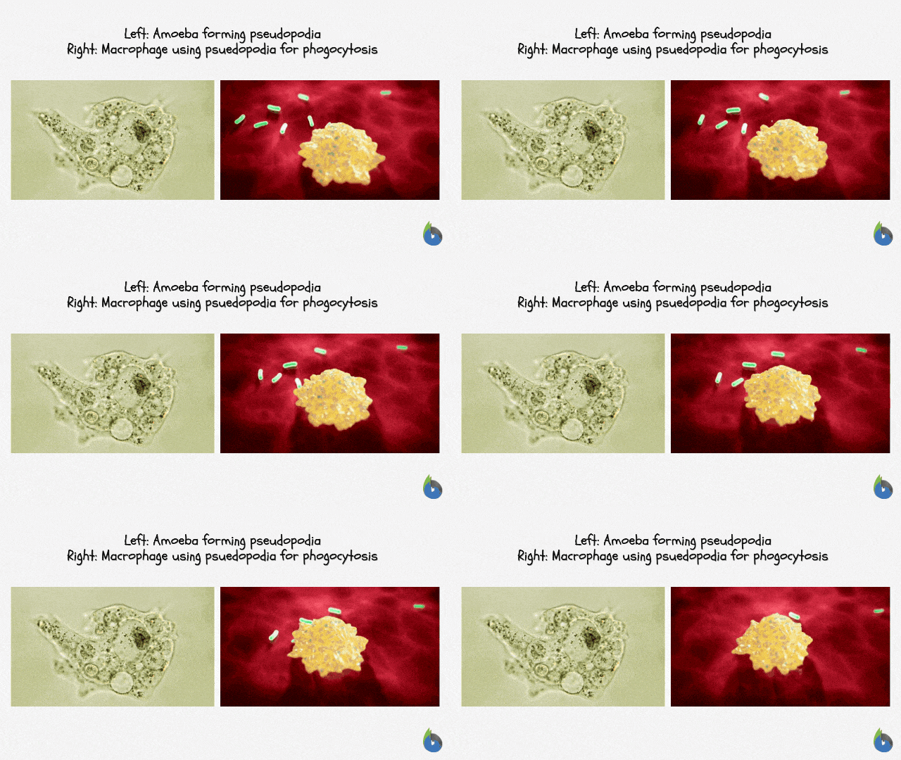
The genus Amoeba (true amoebae) is comprised of single-celled organisms that form pseudopodia. Members of this genus make use of these projections for locomotion and food ingestion. Through them, the amoebas are able to move away from an environment with harsh conditions. This is in addition to other vital mechanisms such as cyst formation and osmoregulation by way of their contractile vacuoles.
Apart from the genus Amoeba, other protists that use pseudopod for analogous functions are the genera Entamoeba and Naegleria. These are medically-important protists as they cause diseases in humans. Entameoba histolytica, for instance, is a pseudopod-forming species that can cause amoebic dysentery. Another is Naegleria fowleri. It is an opportunistic parasite. It is commonly known as the brain-eating amoeba. This species is actually an amoeboflagellate that can enter a human host via the nostrils and then reach the brain tissue to feed on it.
Other amoeboid cells that form pseudopodia are the phagocytic cells of humans. White blood cells, for instance, are cells responsible for the immune response of the body. They move and engulf foreign particles by forming pseudopods. They also perform phagocytosis to clear the body off unwanted cellular debris. The human mesenchymal stem cells are another example. They are migratory cells that form pseudopodia for locomotion.
Try to answer the quiz below to check what you have learned so far about pseudopodia.
See also
References
- Tang, D. D. (2017). “The roles and regulation of the actin cytoskeleton, intermediate filaments and microtubules in smooth muscle cell migration”. Respiratory Research. 18: 54. https://doi.org/10.1186/s12931-017-0544-7
- Etienne-Manneville, S. (2004). “Actin and Microtubules in Cell Motility: Which One is in Control?”. Traffic. 5: 470–77.
- Pseudopodia. (2019). Retrieved from Microworld website: https://www.arcella.nl/2421-2/
- Patterson, D. J. (n.d.). “Amoebae: Protists Which Move and Feed Using Pseudopodia”. Tree of Life Web Project. Retrieved from https://en.wikipedia.org/wiki/Tree-of-Life-Web-Project
- Bosgraaf, L. & Van Haastert, P. J. M. (2009). “The Ordered Extension of Pseudopodia by Amoeboid Cells in the Absence of External Cues”. PLoS One. 4 (4): 626–634. doi:10.1371/journal.pone.0005253.
- Phagocytosis. (2019). Retrieved from Gsu.edu website: http://hyperphysics.phy-astr.gsu.edu/hbase/Biology/phago.html
- Rosales, C., & Uribe-Querol, E. (2017). Phagocytosis: A Fundamental Process in Immunity. BioMed Research International, 2017, 1–18. https://doi.org/10.1155/2017/9042851
© Biology Online. Content provided and moderated by Biology Online Editors

