Residual volume
n., plural: residual volumes
[ɹɪˈzɪ.d͡ʒu.əl/ ˈvɑ.ljum]
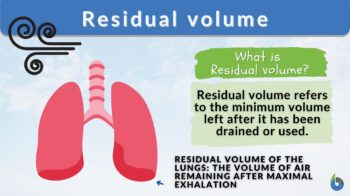
Definition: The minimum volume (level or quantity) left in a bodily organ or environmental reservoir after it has been drained or used
Table of Contents
Residual volume is a term that is most often seen in lung physiology where it is defined as the amount of air remaining in the lungs after maximal exhalation. Apart from lung physiology though, residual volume can also be used similarly in other biological organs.
Here are the biological organs that use the phrase “residual volume” to pertain to the idea of a minimum volume remaining after drainage:
- Lungs: the lung air volume that is not exhaled out even after forceful expiration is known as the residual volume of the lungs. Residual volume air is required to keep the lungs open and inflated at all times. Normally residual volume ranges between 1-1.2 liters.
- Bladder: a residual volume refers to the amount of urine that remains in the bladder after urination, which when atypically high could indicate incomplete bladder emptying
- Gastric Residual Volume: this term is used to describe the volume of liquid or feed remaining in the stomach after a certain period of time, such as after digestion, or during a gastric emptying test.
- Gallbladder: the “residual volume”, in this sense, refers to the amount of bile or gallstones left in the gallbladder after a gallbladder contraction or gallstone passage.
In other sciences, such as chemistry and environmental science, the term is also used. In particular, residual volume in chromatography refers to the portion of a sample that remains on the stationary phase after elution. In environmental science, the residual volume represents the minimum water level or quantity left in a reservoir or water body after it has been drained or used.
In the next sections, though, we will focus more on the definition of residual volume in the lungs, where it is of common use.
Residual Volume Definition
Residual volume is the term used to describe the air volume that remains in the lungs post-forceful expiration. Indirectly, this term also defines or specifies the air volume that can not be expelled from the lungs and is essential for keeping the alveoli open all the time. Thus, it can be interpreted that some amount of air is always present in the lungs to keep it inflated and prevent its collapse. The amount of air that is residual volume remains unaffected by the amount of air present at the time of forceful and maximum expiration. Generally, the residual volume ranges from 1-1.2 liter, however, it is affected by various factors, namely, age, height, gender, level of physical activity, and weight of the person.
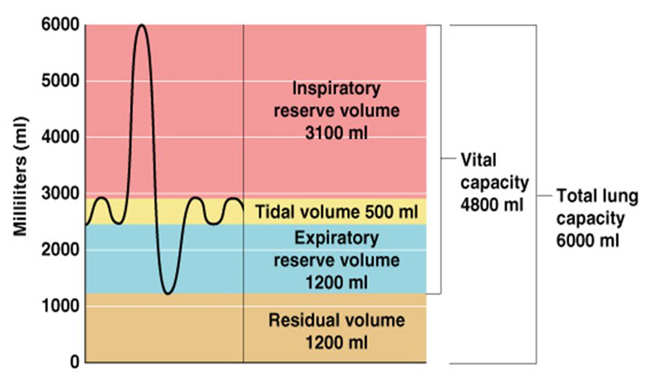
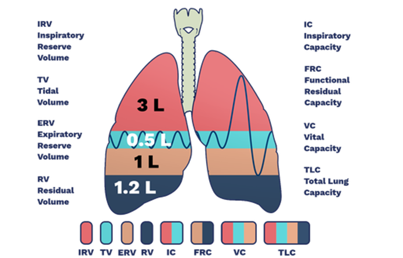
Both the total lung capacity (TLC) and the functional residual capacity (FRC) are inclusive of residual volume:
- TLC represents the amount/air volume in the lungs at maximum inspiration
- FRC is the amount/air volume at the end of normal expiration (Figure 1).

Watch this vid about residual volume in the human lungs:
Biology definition:
In general, a residual volume refers to the minimum volume (level or quantity) left (in a bodily organ or environmental reservoir) after it has been drained or used. For instance, the residual volume of the lungs is the volume of gas that remains in the lungs following a maximal expiration.
Synonym: end-expiratory volume (lung physiology)
Abbreviations: RV; Vr
Why Do We Need Residual Volume?
Residual volume ensures that lungs remain inflated at all times, else in the absence of residual volume, the lung tissue would collapse and stick to each other. This would make the breathing process impossible. Thus, residual volume is necessary for the normal breathing process and maintains lung potency even after maximal expiration.
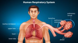
The residual volume of air is used for gaseous exchange between two breaths. Later, the oxygen inspired during respiration replenishes the oxygen-depleted residual air with fresh oxygen and makes it ready for gaseous exchange in the next intermediate phase between two breaths. Also, residual volume is one of the primary parameters that tell about lung health. Hence, residual volume is determined by pulmonary function testing as a reflection of lung physiology and health.
Mechanism
The overall breathing process is complicated, however, it is based upon the basic principle — air flows from a high-pressure region to a low-pressure region. At the physiological resting state, the amount or the volume of the air that enters during inspiration and leave during expiration is referred to as tidal volume (TV).
During tidal inspiration, contraction of the inspiratory muscles of the lungs takes place. This results in an increase in chest volume. This increase in chest volume results in the generation of low intrapleural pressure (Ppl) pressure. The Ppl decreases from (-)5 cm H2O to (-)8 cm H2O.
As a result, the alveolar pressure decreases by 1 cm below the atmospheric pressure. This reduction in alveolar pressure below atmospheric pressure results in the movement of air into the low-pressure alveoli, which creates the residual volume.
Tidal inspiration is an active process whereas expiration is a passive process. During tidal inspiration, the inspiratory muscles contract while in tidal expiration the muscles relax.
NOTE IT!
The maximum amount of air that is expelled from the lungs after forceful expiration is known as vital capacity. Vital capacity is a combination of tidal volume and residual volume.
Difference Between Tidal volume and Residual volume
Tidal volume and residual volume are two separate terms that can be differentiated as specified in the table below (Table 1).
Table 1: Tidal Volume vs Residual Volume | |
|---|---|
| Tidal Volume TV | Residual Volume RV |
| It is the volume of air that is inhaled and exhaled during respiration when the person is at the physiologic resting state or normally breathing | Residual volume is the volume or amount of air which remains or is left in the lungs even after forceful expiration |
| Normally, it is around 0.5 lite (500 ml) | Normally it is between 1-1.2 liters (1000ml- 1200 ml) |
Data Source: Dr. Amita Joshi of Biology Online
Measuring The Residual Volume
None of the pulmonary function tests can measure residual volume directly. The residual volume of the lungs is measured indirectly. It is the only lung volume that is measured indirectly. Prior to estimating residual volume, other lung volumes and capacity have to be estimated. To estimate residual volume, the first functional residual capacity (FRC) is to be calculated. The measurement of FRC can be carried out using either of the following lung function testing methods-
Helium Dilution Test
For measuring lung volumes like FRC, a helium dilution test is used. In this test, the candidate inhales the specific volume of air (V1) that contains a known amount of helium gas at the tidal expiration (FHe1). A spirometer is used to estimate the fraction of helium after equilibrium in the lungs (FHe2).
Hence,
FRC= V1*(FHe1-FHe2)/FHe2
This method is also one of the main tests for measuring static lung volumes.
Nitrogen Washout
This lung function test is also known as Fowler’s method. We all are aware that the air we breathe contains 78% nitrogen. In this test, the candidate breathes through a two-way valve wherein the inspiration valve is connected to the 100% oxygen terminal while the expiration valve is connected to the spirometer.
The spirometer, in this test, is used to estimate the amount/volume of exhaled air and the amount of nitrogen present in each breath. When for three consecutive breaths, the amount of nitrogen falls below 1.5%, the test is considered to be complete. The total amount of nitrogen exhaled is considered to be equivalent to the total amount of nitrogen present. Thus, FRC can be calculated using the following formula:
FRC=Vol. exhaled * Concentration of exhaled N2/ Concentration of initial alveolar N2
Body Plethysmography
This method is based upon Boyle’s law of gases wherein, at a constant temperature, the pressure of a gas is inversely proportional to the volume of gas. Accordingly, the product of the volume of a gas of given mass and its pressure remains constant, i.e.,
P1V1= P2 C2
Given this law, the candidate is placed in a chamber and breathes through a spirometer, to measure the change in volume and pressure. After recording the tidal breathing pattern, the spirometer is closed and the candidate has to make an effort to breathe through it. The changes in pressure-induced due to the closure of the spirometer are recorded. The volume of the thoracic cavity is calculated from the pressure changes that occur during exhalation. This test is one of the reliable and accurate, however expensive tests to estimate FRC.
Once FRC is estimated using any of the above three pulmonary function tests, a spirometer is used to estimate the vital capacity (VC) and expiratory reserve volume (ERV).
For calculating the total lung capacity (TLC) and Tidal Volume (TV) the formulas below are used:
Residual Volume (RV)= FRC- ERV
TLC = VC+ RV
TLC has been found to decrease in patients with chest wall abnormalities whereas TLC increases in patients with obstructive lung disease. To have a clear understanding of all the lung capacities and static lung volumes (RV, TLC, FRC), refer to Figure 2.

The Effect Of Disease On Residual Volume
Obstructive Lung Diseases
Obstructive Lung Diseases are inflammatory disorder wherein expiratory flow obstruction, collapsible airways, and air trapping is seen in the respiratory system. These characteristics lead to an increase in residual volume as compared to healthy lungs.
The most common obstructive lung diseases are asthma, bronchiectasis, and chronic obstructive pulmonary disease (COPD). In Obstructive Lung Diseases, bronchoconstriction, increased mucus production, reduced elasticity of lungs, and inflammation result in elevated airway resistance and closure of small airways.
Closure of airways results in elevated levels of trapped air at the end of expiration, which is referred to as air trapping. Air trapping also results in lung hyperinflation, because of which patients of Obstructive Lung Diseases exhibit higher residual volume (RV), FRC, and TLC.
Patients with Obstructive Lung Disease exhibit higher FRC when estimated with Body plethysmography. An increase in RV is a good indicator of probable Obstructive Lung Disease and pulmonary function. Asthmatic patients exhibit low forced expiratory volume which is the amount of air that can be expelled in a specified time (t sec) in forced breathing.
Restrictive Lung Diseases
Patients with Restrictive Lung Disease exhibit restriction in pulmonary expansion which can be an outcome of pulmonary fibrosis, sarcoidosis, or, processes like obesity and kyphosis. Restricted pulmonary expansion results in a reduction in airflow towards the lungs, created by a negative air pressure gradient. Reduced airflow in the lungs results in decreased lung ventilation and volume. Thus, restrictive lung diseases are characterized by decreased residual volume, TLC, and FRC. It has been observed that in obese patients higher BMI is directly correlated with reduced RV, TLC, and FRC.
Drowning
An important application of RV is for the identification of victims of drowning in post-mortem autopsy. Residual volume air in the lungs can be completely eliminated only in case when it is replaced by another substance. In the case of drowning, the residual volume of air is replaced by the water. Hence, during the autopsy, the lungs are submerged in water, if the lungs don’t float i.e., the lungs sink then it is an indication of death by drowning wherein the victim has inhaled a large amount of water which has replaced the residual volume air and hence, lungs sink. However, in cases wherein the lungs float, the victim might have died before getting into the water.
Recap Questions
- Where is residual air located in the lungs?
a. Trachea
b. Bronchi
c. Alveoli
d. Nostrils
Answer: Option c. Residual air is the air that remains in the alveoli of the lungs even after forceful expiration.
- Which of the following describes total lung capacity?
a. Inspiratory reserve volume (IRV) + Residual Volume (RV)
b. Inspiratory capacity+ Residual Volume (RV)
c. Expiratory reserve volume (ERV) + Residual Volume (RV)
d. Vital capacity (VC) + Residual Volume (RV)
Answer: Option d. Vital capacity is the maximum amount of air expelled from the lungs after forceful inhalation. Residual volume is the volume of air that is expelled after forceful expiration. Hence, total lung capacity is the sum of vital capacity and residual volume.
- If arranged chronologically, which of the following lung volume is the least of all?
a. Tidal volume
b. Vital capacity
c. Residual volume
d. Expiratory reserve volume
Answer: Option a. Reference values for tidal volume is 500 ml whereas residual volume is around 1200 ml. Expiratory reserve volume is 1100 ml and vital capacity is around 4800 ml. Hence, tidal volume is the least of all the lung volumes.
Take the Residual Volume – Biology Quiz!
References
- Flesch, J. D., & Dine, C. J. (2012). Lung volumes: measurement, clinical use, and coding. Chest, 142(2), 506–510. https://doi.org/10.1378/chest.11-2964
- Kwon, O. B., Yeo, C. D., Lee, H. Y., Kang, H. S., Kim, S. K., Kim, J. S., Park, C. K., Lee, S. H., Kim, S. J., & Kim, J. W. (2021). The Value of Residual Volume/Total Lung Capacity as an Indicator for Predicting Postoperative Lung Function in Non-Small Lung Cancer. Journal of clinical medicine, 10(18), 4159. https://doi.org/10.3390/jcm10184159
- Lofrese, J. J., Tupper, C., Denault, D., & Lappin, S. L. (2023). Physiology, Residual Volume. In StatPearls. StatPearls Publishing.
- Shin, T. R., Oh, Y. M., Park, J. H., Lee, K. S., Oh, S., Kang, D. R., Sheen, S., Seo, J. B., Yoo, K. H., Lee, J. H., Kim, T. H., Lim, S. Y., Yoon, H. I., Rhee, C. K., Choe, K. H., Lee, J. S., & Lee, S. D. (2015). The Prognostic Value of Residual Volume/Total Lung Capacity in Patients with Chronic Obstructive Pulmonary Disease. Journal of Korean medical science, 30(10), 1459–1465. https://doi.org/10.3346/jkms.2015.30.10.1459
©BiologyOnline.com. Content provided and moderated by Biology Online Editors.


