Genetic Mutations
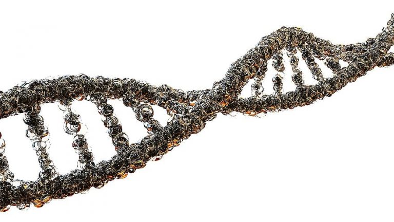
Schematic diagram of DNA
Table of Contents
Reviewed by: Mary Anne Clark, Ph.D.
Genetic Mutations
Genetic mutations are inherited variations in an organism’s appearance or function. At the molecular level, genetic mutations are the consequence of a change in the DNA sequence of a single gene. In protein-coding genes, some changes in DNA sequence alter or even obliterate the function of the encoded protein. Others may change the amino acid sequence without damaging the protein, and still others may leave the amino acid sequence unchanged. The critical factor is whether or not the mutant protein refolds into a nonfunctional form. Mutations may occur either because of a replication error or as a consequence of altering the DNA by radiation or chemical damage.
Single Nucleotide Polymorphisms
Single-nucleotide polymorphisms, also called point mutations, are changes to a single base in the coding sequence of a gene. For example, the first 30 bases in the coding sequence of the human beta-globin gene are written below, broken into three-base codons and followed by the encoded amino acids of beta-globin.
![]()
A single base change may have one of several consequences for the encoded amino acid sequence. One feature of the genetic code is that it is redundant, that is, most amino acids are specified by two or more codons, and some have as many as six! For example, CTG, the fourth codon in the sequence above, specifies the amino acid leucine (L). Changes in the third base of the codon will still produce a leucine codon. TTA and TTG also encode leucine, so a single base change from CTG to TTG will also leave the amino acid unchanged. A mutation like this, that changes the DNA codon, but does not change the amino acid, is called a silent mutation.
Mutations that result in substituting one amino acid for another are called missense mutations. One of the most well-known of the missense mutations is the one that produces sickle-cell disease, a change in the first GAG codon in the sequence above to GTG. This mutation results in trading the normal amino acid glutamate (E) for the amino acid valine (V). Glutamate is negatively charged and valine is a large water-insoluble and uncharged amino acid. It is this change that leads to the hemoglobin molecules sticking together to form long spiky structures that deform the red blood cells. On the brighter side, it also confers resistance to infection with the malaria parasite. Other missense mutations may change the amino acid, but to a codon that specifies an amino acid with similar properties, e.g. changing CTG to GTG, which encodes valine. Both leucine and valine are large water-insoluble amino acids, so changes like that may be relatively inconsequential.
A much more damaging kind of mutation is to introduce an early stop codon. For example in some cases of beta-thalassemia, a form of anemia, the codon CAG for the amino acid glutamine at position 39 in the protein is replaced by the stop codon TAG, terminating the protein at a third of its normal length, and making it completely nonfunctional. Mutations of this type are called nonsense mutations. Other early termination mutations in the beta-globin gene have also been associated with beta-thalassemia.
Temperature-sensitive Mutations
Some missense mutations can produce a protein that folds normally at the normal temperature for the organism but becomes unstable at higher temperatures. An excellent example of a temperature-sensitive mutation is seen in Siamese cats. The gene affected is the gene for the enzyme tyrosinase, which catalyzes the first step in the synthesis of the pigment melanin from the amino acid tyrosine. The normal protein is over 500 amino acids long but in Siamese cats the amino acid glycine (G) at position 302 is changed to arginine (R). Siamese kittens are unpigmented when they are born, because of their exposure to the warmth of their mother’s body, but the ears, nose, tail, and feet, which are cooler than the core of the body, darken as they get older. In these regions, the enzyme is working, but over the warmer parts of the body, it does not.
Indels
Indels are mutations resulting from the insertion or deletion of one or more nucleotides in a DNA sequence. Unless the indel involves an entire codon or a segment of DNA that is some multiple of three nucleotides, the mutation produces a shift in the reading frame when the protein is translated. An example of a shift in the reading frame is the removal of the second “b” from the sentence “Bob can eat the egg” to transform it into “Boc ane att hee gg.” Although only a single element has been lost from the sentence, it is now devoid of meaning. Indels frequently produce such frameshift mutations, which make their encoded proteins equally useless. A common consequence of frameshifts is the generation of stop codons so that the proteins are both meaningless and short!
Not all indels produce frameshifts, but even the loss of a single amino acid from a protein can be deleterious. For example, the most commonly found mutation in cystic fibrosis is an indel that leads to the loss of the amino acid phenylalanine #508 in CFTR, cystic fibrosis transmembrane conductance regulator protein. This protein is famously large at nearly 1500 amino acids, but the loss of that single phenylalanine significantly reduces its ability to do its normal job of transporting chloride ions across the cell membrane and helping to regulate tissue fluids.
Trinucleotide Repeat Expansions
Some proteins have a run of the same amino acid repeated more than a dozen times due to repetition of the same or similar codons Since the DNA in repeated regions of paired chromosomes looks very similar, it is subject to crossing-over errors which can result in either shortening or lengthening the run of amino acids. Either can lead to misfolding of the protein, but several genetic disorders are due to expansions of the repeated region. One of these trinucleotide repeat disorders is Huntington’s Disease, which is associated with an increase in the length of the CAG repeat region in the Huntingtin protein from two dozen glutamines (Q), to 40 or more. Why the expanded version of the protein progressively damages neural tissue is not well understood. Other trinucleotide repeat disorders include myotonic dystrophy and fragile-X syndrome.
Gene Duplication
At the margin of the boundary between single-gene mutations and chromosome level mutations are gene duplications, in which one or a few genes are tandemly duplicated. After duplication, the “extra” gene copy is free to accumulate new mutations and experiment with new functions.
Duplicated genes are an important source of developmental regulation and the production of new genes and gene families. Using the beta globins again as an example, the beta globins themselves are the product of gene duplications, since both the beta and alpha globins are historically related to myoglobin, and the three genes are now on separate chromosomes. In humans, the “normal” adult beta-globin itself is produced from one of a cluster of beta-globin genes on human Chromosome 11. There are five functional genes plus a pseudogene, which is no longer functional in humans. Each of the five genes is used at a different developmental stage. The epsilon globin produces the beta chain of embryonic hemoglobin. The two gamma chains, gamma-a and gamma-b, produce the beta chains of fetal hemoglobin. Finally, the beta and delta chains take over at birth and become the postnatal beta chains, with the delta chain as a minor variant in human hemoglobin.
Another intriguing example of the result of gene duplication is in the red and green color receptor proteins in humans and the great apes. Both are on the X chromosome, side by side. In some monkeys, the X-linked red and green genes are alleles in females. Most mammals have only the green gene. The red and green opsins are 96% identical, but the amino acid differences between them are sufficient to produce two different maximum absorption profiles in the retina, the red opsin at 560nm and the green opsin at 530 nm. In some humans, there are even additional copies of the green gene. One wonders what new colors humans may be able to see in the future.
Polyploidy is responsible for the creation of thousands of species in today’s planet and will continue to do so. It is also responsible for increasing genetic diversity and producing species showing an increase in size, vigor, and increased resistance to disease. Know more about it in the next tutorial. It also includes topic on mutation frequency.
 | GENETIC MUTATIONS – MODIFIED TRUE OR FALSE A modified true-or-false quiz meant to test the students’ comprehension of the principles and vocabulary in genetic mutations. Apart from identifying which items are correct and incorrect, the students also have to correct the false statements. Subjects: Genetics & Evolution |
You will also like...
Plant Tissues
Plant organs are comprised of tissues working together for a common function. The different types of plant tissues are m..
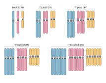
Polyploidy
Humans are diploid creatures. This means that for every chromosome in the body, there is another one to match it. Howeve..

Sigmund Freud and Carl Gustav Jung
In this tutorial, the works of Carl Gustav Jung and Sigmund Freud are described. Both of them actively pursued the way h..

Biosecurity and Biocontrol
This lesson explores the impact of biosecurity threats, and why they need to be identified and managed. Examples to incl..
Effect of Chemicals on Growth & Development in Organisms
Plants and animals need elements, such as nitrogen, phosphorus, potassium, and magnesium for proper growth and developme..
Cell Respiration
Cell respiration is the process of creating ATP. It is "respiration" because it utilizes oxygen. Know the different stag..
