Neural Control Mechanisms

Neurons generate electric signals that pass from one end of the neuron to another
Table of Contents
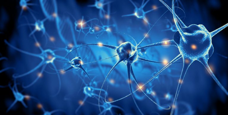
Nerve cells called neurons generate electric signals that pass from one end of the cell to another and release chemical messengers called neurotransmitters to communicate with other cells.
Structure
A neuron has: (1) a cell body containing the cell organelles, (2) dendrites, branched outgrowths from the cell body that receive inputs over its vast surface area, (3) an axon, a single long process that extends from the cell body to its target cells, (4) an axon terminal which releases neurotransmitters that diffuse through extracellular space to trigger cells opposite the terminal.
A nerve fiber is a single axon while a nerve is a bundle of axons bound together by connective tissue.
Axons of some neurons are covered by myelin, a layer of plasma membranes with supporting cells that are called glial cells in CNS and Schwann cells in the peripheral nervous system. The spaces between adjacent sections of myelin where the axon is exposed to extracellular fluid are called nodes of Ranvier. Myelin speeds up the conduction of electric signals.
Glial Cells
Glial cells physically and metabolically support neurons. Oligodendroglia form the myelin covering of CNS axons. Astroglia regulates the composition of extracellular fluid in CNS. Microglia perform immune functions.
Functional Classes of Neurons
3 types:
- Afferent neurons that have sensory receptors at their ends and convey signals from tissues and organs into CNS
- Efferent neurons that transmit signals from CNS to effector cells
- Interneurons that connect neurons within CNS.
The junction between two neurons, where one neuron alters the activity of another (via a neurotransmitter) is called a synapse. A neuron conducting signals toward a synapse is called a presynaptic neuron while a neuron conducting signals away from a synapse is a postsynaptic neuron.
Neural Growth and Regeneration
The development of neurons is guided by neurotropic (neurogrowth) factors. Neurons outside the CNS can repair themselves but neurons within the CNS cannot.
Membrane Potentials
The difference in the amount of charge between two points is called a potential difference and its unit of measurement is volt. This difference tends to make the charge low, producing an electric current. The material through which it is flowing obstructs the current and this is called resistance. Ohm’s law gives the relationship
I = E/R
where:
I = electric current
E = electric potential
R = resistance
Materials with high resistance are called insulators, and those with low resistance are called conductors. Water with dissolved ions (electrolytes) is a good conductor while lipids are insulators. Intra- and extracellular fluids have numerous ions and are therefore conductors while the plasma membrane separating them is an insulator.
Resting membrane potential
The potential difference across the plasma membrane of a cell under resting conditions inside of the cell is negatively charged with respect to outside. The magnitude of the potential is determined by (1) differences in specific ion concentrations in intra- and extracellular fluids, and (2) differences in membrane permeabilities to different ions as a function of the number of open ion channels for these ions. Na+ and K+ play the most important roles in generating the resting membrane potential. Nat is greater outside while K+ is greater inside the cell. K+ moves out of the cell and Na+ moves into the cell down their concentration gradients but an intracellular concentration of these two ions is kept constant by an active transport system that pumps Na+ out of the cell and K+ into it. However, the pump brings out 3 Na+ for every 2 K+ it pumps in, making inside of the cell negative.
Graded and action potentials
Graded potentials are changes in membrane potential confined to a small region of the plasma membrane. The magnitude of these potentials is related to the magnitude of the initiating stimulus. They initiate a signal. Action potentials are large, rapid alterations in the membrane potential. Membranes capable of producing action potentials are called excitable membranes. Examples are membranes in nerve and muscle cells.
Ionic basis of action potential
During an action potential, voltage-gated Na+ channels open and allow a large influx of Na+ ions into the cell, making inside of the cell less negative and this is called depolarization. The membrane starts returning rapidly to the resting membrane potential because Na+ channels close, voltage-gated K+ channels open, K+ moves out and this is called repolarization. However, so much K+ moves out that inside of the cell becomes more negative than the original resting membrane potential and this is called hyperpolarization. In some cells, Ca2+ channels serve the same function as Na+ channels. Local anesthetics block the Na+ channels and prevent an action potential.
Threshold and all-or-none response
The potential at which a membrane is depolarized to generate an action potential is called the threshold potential and stimulus that is strong enough to depolarize the membrane is called a threshold stimulus.
A stimulus of more than threshold magnitude also elicits an action potential of the same amplitude as that caused by a threshold stimulus. This is because once the threshold is reached membrane events are no longer dependent upon the stimulus strength. Therefore, action potentials occur maximally or do not occur at all and this is called an all-or-none response. This is why a single action potential cannot convey information about the magnitude of the stimulus that initiated it
Refractory periods
The period after an action potential when a second stimulus will, not produce a second action potential is called an absolute refractory period. It occurs because once the voltage-gated Na+ channels close, the membrane needs to repolarize before the channels can open once again. Following the absolute refractory period, there is an interval during which a second action potential can be produced only is the stimulus strength is greater than usual. This is called relative refractory period and is a result of hyperpolarization.
Initiation of action potential
The initial depolarization in afferent neurons is achieved by either a graded potential called receptor potential in the receptors or by a spontaneous change in the neuron membrane potential called pacemaker potential.
Action potential propagation
Since a neuron is a long cell, it gets depolarized part by part and not all at once. The area of the membrane that gets depolarized has a difference in potential with the adjacent area of the membrane that is still at resting potential causing a local current. This current then depolarizes the adjacent resting membrane and a new action potential is generated there and so on. Because depolarization of an area is followed by a refractory period, the action potential moves unidirectionally. The velocity of action potential propagation is positively correlated with fiber diameter because a larger fiber offers less resistance. Myelin sheath, being an insulator prevents the flow of ions between intra- and extracellular compartments. Therefore, action potentials occur only at the non-insulated nodes of Ranvier and this jump of action potentials from one node to another is called saltatory conduction. By preventing leakage of charge, myelin increases the speed of propagation, enabling axons to be thinner.
Synapses
A synapse is a junction between two neurons, where the electrical activity in the presynaptic neuron influences the electrical activity in the postsynaptic neuron. The influence can be either excitatory or inhibitory. If many presynaptic cells affect a single postsynaptic cell it is called convergence and allows information from many sources to influence the activity of one cell. If a single presynaptic cell affects many postsynaptic cells it is called divergence and allows one information source to affect multiple pathways.
Functional anatomy of synapses
At electric synapses, the pre- and postsynaptic cells are joined by gap junctions, allowing action potentials to flow directly across the junction. Such synapses are rare.
At chemical synapses, the axon of the presynaptic neuron ends in a swelling called the axon terminal and an extracellular space called the synaptic cleft separates the pre- and postsynaptic neurons, preventing direct propagation of current between them. Signals are transmitted across the synaptic cleft by a chemical messenger – a neurotransmitter – released from the presynaptic axon terminal and bound by receptors at the postsynaptic cell. Most chemical synapses operate in only direction.
Neurotransmitters in axon terminals are stored in membrane-bound synaptic vesicles that are docked at the synaptic membrane. When an action potential depolarizes the axon terminal, voltage-gated Ca2+ channels in the membrane open, and Ca2+ diffuses from extracellular space into the axon terminal. The Ca2+ induce reactions that allow the vesicles to fuse with the plasma membrane and liberate their contents into the synaptic cleft by exocytosis.
Excitatory chemical synapses
The activated receptor on the postsynaptic membrane opens Na+ channels. There is a net movement of Na+ ions into the cell, resulting in depolarization. This potential change in the postsynaptic neuron is called an excitatory postsynaptic potential (EPSP). It is a graded potential.
Inhibitory chemical synapses
The activated receptor on the postsynaptic membrane opens Cl- channels. There is a net movement of Cl- ions into the cell, resulting in hyperpolarization. The potential change in the postsynaptic neuron is called an inhibitory postsynaptic potential (IPSP). It is a graded potential.
Activation of the postsynaptic cell
In most neurons, one EPSP is not enough to cross the threshold in the postsynaptic neuron and only the combined effects of many excitatory synapses can initiate an action potential. If a number of EPSPs arriving at different times create a depolarization it is called a temporal summation. If a number of EPSPs arriving at different locations create a depolarization, it is called a spatial summation. IPSPs also show similar summations but the effect is a hyperpolarization.
Neurotransmitters and Neuromodulators
Neuromodulators modify the postsynaptic cell’s response to neurotransmitters or change the presynaptic cell’s synthesis, release or metabolism of the neurotransmitter.
Acetylcholine (Ach)
Major neurotransmitter. Fibers that release ACh are called cholinergic fibers. Acetylcholine is degraded by the enzyme, acetylcholinesterase.
Biogenic amines
Biogenic amines are neurotransmitters containing an amino group. Catecholamines such as dopamine, norepinephrine and epinephrine, serotonin. Nerve fibers that release epinephrine and norepinephrine are called adrenergic and noradrenergic fibers respectively.
Amino acid neurotransmitters
Amino acid neurotransmitters are the most prevalent neurotransmitters in CNS. Glutamate, aspartate GABA (gamma-aminobutyric acid), glycine,
Neuropeptides
Neuropeptides are composed of two or more amino acids. Neurons releasing neuropeptides are called peptidergic. Beta-endorphin, dynorphin, enkephalins.
Nitric oxide, ATP, adenine also act as neurotransmitters.
Neuroeffector communication
Many neurons of the peripheral nervous system end at neuroeffector junctions on muscle and gland cells. Neurotransmitters released by these efferent neurons then activate the target cell.
Structure of the Nervous System
A group of nerve fibers traveling together in the CNS is called a pathway or tract and if it joins the left and the right halves, it is called a commissure.
Information in CNS passes along two types of pathways:
- Long neural pathways in which neurons with long axons carry information directly between the brain and spinal cord or between different regions of the brain. There is no alteration in the transmitted information.
- Multineuronal or multisynaptic pathway. Made up of many neurons or synapses. New information can be integrated into the transmitted information.
Cell bodies of neurons having similar function cluster together and such clusters are called ganglia in the peripheral nervous system and nuclei in the CNS.
CNS: spinal cord
The spinal cord lies within the vertebral column. The central gray matter is composed of interneurons, cell bodies, dendrites, and glial cells. This is surrounded by white matter composed of myelinated axons of interneurons. The fiber tracts either descend to relay information from the brain or ascend to relay information to the brain or transmit information across different levels of the spinal cord.
Afferent fibers enter from the peripheral system enter on the dorsal side of the cord via dorsal roots and form the dorsal root ganglia. Efferent fibers leave the cord on the ventral side via ventral roots. Dorsal and ventral roots from the same level combine to form a spinal nerve outside the cord, one on each side. 31 pairs of spinal nerves are designated by 4 levels of exit – cervical (8), thoracic (12), lumbar (5), and sacral (5).
CNS: brain
Brainstem: Consists of midbrain, pons and medulla oblongata. It contains the reticular formation, a bundle of axons that is involved in motor functions, cardiovascular and respiratory control, etc.
Cerebellum: An important center for coordinating movements and for controlling balance and posture.
Forebrain: The larger component of the forebrain, the cerebrum consists of the right and left cerebral hemispheres that have an outer shell of gray matter the cerebral cortex. Each hemisphere is divided into 4 lobes: frontal, parietal, occipital and temporal. The cortex is the most complex integrating area. The central core of the brain is formed by the diencephalon consisting of the thalamus – a collection of several large nuclei, and the hypothalamus – the master command center for neural and endocrine coordination.
Peripheral nervous system
Transmits signals between the CNS and receptors/effectors. Consists of 12paairs of cranial nerves that connect with the brain and 31 pairs of spinal nerves that connect with the spinal cord.
The efferent system is further divided into a somatic and an autonomic system.
Somatic nervous system
Innervates skeletal muscles. Consists of myelinated axons without any synapses. The activity of these neurons leads to excitation (contraction) of skeletal muscles and therefore they are called motor neurons. They are never inhibitory.
Autonomic nervous system
Innervates smooth and cardiac muscles. Parallel chains, each with two neurons, connect the CNS and effector cells. The synapse between these two neurons is called the autonomic ganglion, the nerve fibers between the CNS and the ganglion are called pre-ganglionic fibers and those between the ganglion and the effector cells are called post-ganglionic fibers.
Further divided into sympathetic (fight or flight) and parasympathetic (rest and relax) components.
Sympathetic ganglia lie close to the spinal cord while parasympathetic ganglia lie close to the organs. The sympathetic system is arranged to act as a single unit while the parasympathetic system is arranged such that the parts can act independently. The sympathetic system is involved in responses to stress. Many organs and glands receive a dual innervation from both sympathetic and parasympathetic fibers. The two systems generally have opposite effects and work together to regulate a response. Most autonomic responses usually occur without conscious control.
Blood supply, blood-brain barrier and cerebrospinal fluid
The neural tissue of the CNS is covered by 3 membranes called meninges – the outermost dura mater, the middle arachnoid, and the inner pia mater. The space between the pia and the arachnoid, the subarachnoid space, is filled with cerebrospinal fluid (CSF). It acts as a shock absorber for neural tissue.
The brain is highly dependent on a continuous supply of glucose and oxygen via blood. It has little stored glycogen.
The exchange of substances between the blood and extracellular fluid in CNS is highly restricted via a complex group of blood-brain barrier mechanisms. The CSF and the extracellular fluid in the brain are in diffusion equilibrium with each other but maintain a difference with the blood.
You will also like...

Ecology & Biodiversity: New Zealand Flora & Fauna
New Zealand is known for its unique biodiversity, caused by its remarkable geography and geologic history. Breaking away..

Arthropods
The arthropods were assumed to be the first taxon of species to possess jointed limbs and exoskeleton, exhibit more adva..

Stems
Stems primarily provide plants structural support. This tutorial includes lectures on the external form of a woody twig ..

Types and Causes of Brain Damage
This tutorial describes the different types and causes of brain damage. Find out how genetics, physical injury, lack of ..

Community Patterns
Learn about community patterns and the ecological factors influencing these patterns. Revisit some of the ecosystems you..

Still Water Animals
Animals living in aquatic habitats have diversified and evolved through time. They eventually occupy ecological niches a..
